

Measurement of tissue scattering properties using multi-diameter single fiber reflectance spectroscopy: in silico sensitivity analysis
- PMID: 22076275
- PMCID: PMC3207383
- DOI: 10.1364/BOE.2.003150
Measurement of tissue scattering properties using multi-diameter single fiber reflectance spectroscopy: in silico sensitivity analysis
Abstract
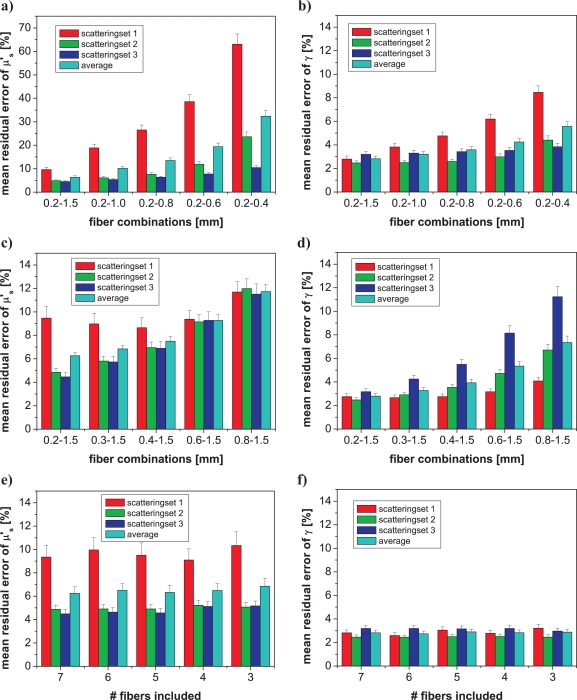
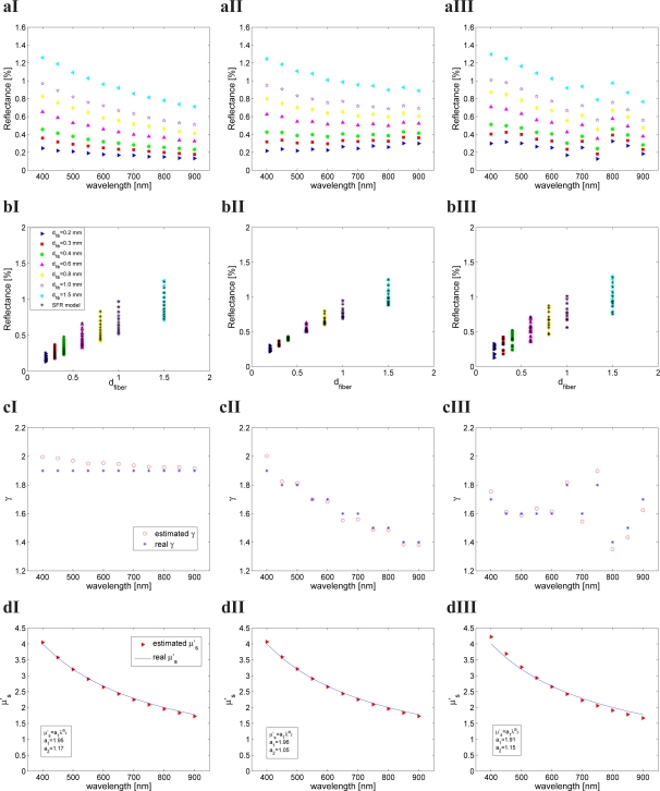
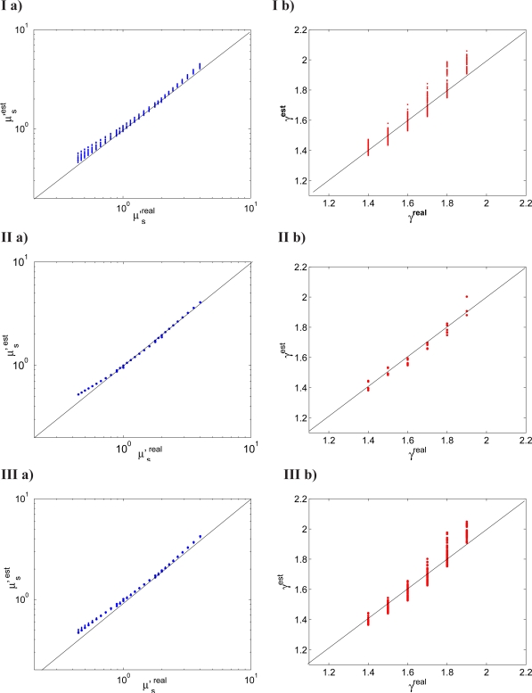
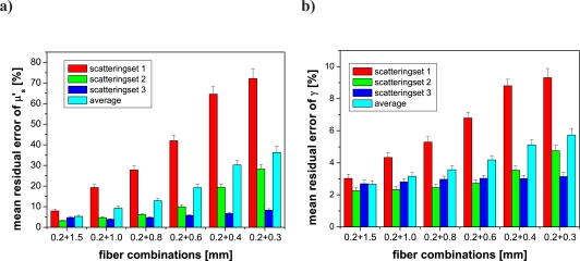
Multiple diameter single fiber reflectance (MDSFR) measurements of turbid media can be used to determine the reduced scattering coefficient (μ'(s)) and a parameter that characterizes the phase function (γ). The MDSFR method utilizes a semi-empirical model that expresses the collected single fiber reflectance intensity as a function of fiber diameter (d(fiber)), μ'(s), and γ. This study investigated the sensitivity of the MDSFR estimates of μ'(s) and γ to the choice of fiber diameters and spectral information incorporated into the fitting procedure. The fit algorithm was tested using Monte Carlo simulations of single fiber reflectance intensities that investigated biologically relevant ranges of scattering properties (μ'(s) ∈ [0.4 - 4]mm(-1)) and phase functions (γ ∈ [1.4 - 1.9]) and for multiple fiber diameters (d(fiber) ∈ [0.2 - 1.5] mm). MDSFR analysis yielded accurate estimates of μ'(s) and γ over the wide range of scattering combinations; parameter accuracy was shown to be sensitive to the range of fiber diameters included in the analysis, but not to the number of intermediate fibers. Moreover, accurate parameter estimates were obtained without a priori knowledge about the spectral shape of γ. Observations were used to develop heuristic guidelines for the design of clinically applicable MDSFR probes.
Keywords: (170.1470) Blood or tissue constituent monitoring; (170.3660) Light propagation in tissues; (170.6510) Spectroscopy, tissue diagnostics; (290.7050) Turbid media; (300.6540) Spectroscopy, ultraviolet; (300.6550) Spectroscopy, visible.
Figures
Similar articles
-
In vivo quantification of the scattering properties of tissue using multi-diameter single fiber reflectance spectroscopy.Biomed Opt Express. 2013 Apr 9;4(5):696-708. doi: 10.1364/BOE.4.000696. Print 2013 May 1. Biomed Opt Express. 2013. PMID: 23667786 Free PMC article.
-
Use of a coherent fiber bundle for multi-diameter single fiber reflectance spectroscopy.Biomed Opt Express. 2012 Oct 1;3(10):2452-64. doi: 10.1364/BOE.3.002452. Epub 2012 Sep 12. Biomed Opt Express. 2012. PMID: 23082287 Free PMC article.
-
Semi-empirical model of the effect of scattering on single fiber fluorescence intensity measured on a turbid medium.Biomed Opt Express. 2012 Jan 1;3(1):137-52. doi: 10.1364/BOE.3.000137. Epub 2011 Dec 14. Biomed Opt Express. 2012. PMID: 22254174 Free PMC article.
-
Method for rapid multidiameter single-fiber reflectance and fluorescence spectroscopy through a fiber bundle.J Biomed Opt. 2013 Oct;18(10):107005. doi: 10.1117/1.JBO.18.10.107005. J Biomed Opt. 2013. PMID: 24126725
-
Evaluation of a fiberoptic-based system for measurement of optical properties in highly attenuating turbid media.Biomed Eng Online. 2006 Aug 23;5:49. doi: 10.1186/1475-925X-5-49. Biomed Eng Online. 2006. PMID: 16928274 Free PMC article.
Cited by
-
In vivo quantification of the scattering properties of tissue using multi-diameter single fiber reflectance spectroscopy.Biomed Opt Express. 2013 Apr 9;4(5):696-708. doi: 10.1364/BOE.4.000696. Print 2013 May 1. Biomed Opt Express. 2013. PMID: 23667786 Free PMC article.
-
Extraction of intrinsic fluorescence from single fiber fluorescence measurements on a turbid medium: experimental validation.Biomed Opt Express. 2014 May 22;5(6):1913-25. doi: 10.1364/BOE.5.001913. eCollection 2014 Jun 1. Biomed Opt Express. 2014. PMID: 24940549 Free PMC article.
-
Advances in optics for biotechnology, medicine and surgery.Biomed Opt Express. 2012 Mar 1;3(3):531-2. doi: 10.1364/BOE.3.000531. Epub 2012 Feb 10. Biomed Opt Express. 2012. PMID: 22435099 Free PMC article.
-
Optical detection of field cancerization in the buccal mucosa of patients with esophageal cancer.Clin Transl Gastroenterol. 2018 Apr 30;9(4):152. doi: 10.1038/s41424-018-0023-6. Clin Transl Gastroenterol. 2018. PMID: 29712897 Free PMC article.
-
Limitations of the commonly used simplified laterally uniform optical fiber probe-tissue interface in Monte Carlo simulations of diffuse reflectance.Biomed Opt Express. 2015 Sep 11;6(10):3973-88. doi: 10.1364/BOE.6.003973. eCollection 2015 Oct 1. Biomed Opt Express. 2015. PMID: 26504647 Free PMC article.
References
-
- Drezek R., Guillaud M., Collier T., Boiko I., Malpica A., Macaulay C., Follen M., Richards-Kortum R., “Light scattering from cervical cells throughout neoplastic progression: influence of nuclear morphology, DNA content, and chromatin texture,” J. Biomed. Opt. 8, 7–16 (2003).10.1117/1.1528950 - DOI - PubMed
LinkOut - more resources
Full Text Sources
Research Materials