Review
. 2011 Dec;15(1):2-11.
doi: 10.1038/jidsymp.2011.7.

Clinical, cellular, and molecular aspects in the pathophysiology of rosacea

Affiliations
Review

Clinical, cellular, and molecular aspects in the pathophysiology of rosacea

Martin Steinhoff et al. J Investig Dermatol Symp Proc. 2011 Dec.

Abstract

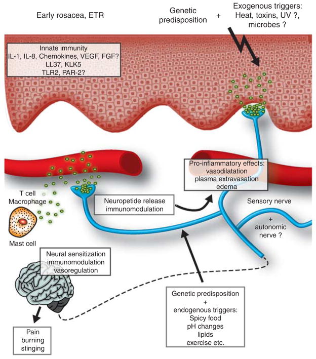
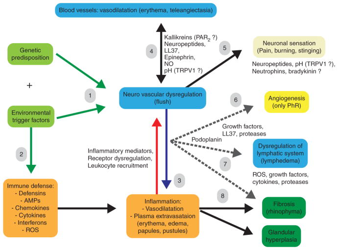
Rosacea is a chronic inflammatory skin disease of unknown etiology. Although described centuries ago, the pathophysiology of this disease is still poorly understood. Epidemiological studies indicate a genetic component, but a rosacea gene has not been identified yet. Four subtypes and several variants of rosacea have been described. It is still unclear whether these subtypes represent a "developmental march" of different stages or are merely part of a syndrome that develops independently but overlaps clinically. Clinical and histopathological characteristics of rosacea make it a fascinating "human disease model" for learning about the connection between the cutaneous vascular, nervous, and immune systems. Innate immune mechanisms and dysregulation of the neurovascular system are involved in rosacea initiation and perpetuation, although the complex network of primary induction and secondary reaction of neuroimmune communication is still unclear. Later, rosacea may result in fibrotic facial changes, suggesting a strong connection between chronic inflammatory processes and skin fibrosis development. This review highlights recent molecular (gene array) and cellular findings and aims to integrate the different body defense mechanisms into a modern concept of rosacea pathophysiology.


Conflict of interest statement

CONFLICT OF INTEREST

The authors state no conflict of interest.

Figures

Figure 1
Figure 1. Relationship between clinical characteristics and gene array profile of rosacea
Three clinical subtypes have been studied by gene array analysis from facial skin biopsies and compared with healthy facial or uninvolved skin (healthy skin, HS). ETR, erythematotelangiectatic rosacea (subtype I, RI); PPR, papulopustular rosacea (subtype II, RII); PhR, phymatous rosacea (subtype III, RIII). (a) ETR is characterized by a prolonged flushing erythema and teleangiectasia. PPR is characterized by papules and, more rarely, pustules, in addition to erythema and telangiectasia. PhR is characterized by fibrotic changes and glandular hyperplasia, less clinically obvious inflammation, and erythema. Although flushing and discomfort are the predominant clinical features, and no papules are visible in ETR, a marked inflammatory infiltrate of CD4+ T lymphocytes, macrophages, and mast cells is visible already in ETR by immunohistochemistry (see also Figure 3). (b) Gene array analysis of biopsies from patients with rosacea (subtypes I–III; n=6–8 patients per subtype) reveals differential regulation of various selective genes among the various subtypes (RI=ETR=cluster II; RII=PPR=cluster III; RIII=PhR=cluster IV) as compared with healthy or uninvolved skin (cluster I). Because certain genes overlap, a developmental “march” from rosacea I to III can be assumed, although it is not always clinically visible (arrows).
Figure 2
Figure 2. Analysis of inflammatory and immune changes in rosacea on the basis of gene array analysis
(a) Biological interpretation of genes upregulated in cluster II (erythematotelangiectatic rosacea, ETR). Of the 221 known genes that clustered in various dominant biological processes, about 20% can be implicated in inflammation and immune defense. A surprising and thus far unacknowledged increase of genes involved in alcohol and lipid metabolism was observed. About 5% of the genes are involved in epidermal activation or development, indicating the involvement of keratinocytes at an early stage of rosacea. (b) Gene array analysis of cytokines and chemokines involved in the pathophysiology of rosacea indicates a role of interleukin (IL)-7, IL26, and various chemokines already in early subtypes of rosacea (see Gerber et al., 2011, for details). RI, ETR; RII, papulopustular rosacea (PPR); RIII, phymatous rosacea (PhR). WAP, whey acidic protein.
Figure 3
Figure 3. Histopathological characteristics of rosacea subtypes (erythematotelangiectatic rosacea (ETR), papulopustular rosacea (PPR), phymatous rosacea (PhR))
All three subtypes were stained for inflammatory and immune markers to determine the inflammatory infiltrate and skin structures involved in inflammation and fibrosis (T cells: CD4, CD8; B cells: CD20, CD79a; Langerhans cells: CD1a; macrophages: CD68; natural killer cells: CD56; neutrophils: CD15, elastase; mesenchymal cells: vimentin (VIM); nerves: PGP9.5, NF200; blood vessels: CD31; lymphatic vessels: podoplanin (Podpl)) in the various subtypes. An increased mast cell infiltrate can also be observed in all subtypes of rosacea (Schwab et al., 2011). (b) Bar=200 μm; (a, g, i, j, lo), bar=100 μm; (c, d, f, h, k, p), bar=50 μm.
Figure 4
Figure 4. Potential role of neurogenic inflammation in the early phase of rosacea
Genetic predisposition, along with exogenous or endogenous trigger factors, stimulates peripheral nerve endings of the skin. Central transmission of neuronal activation leads to facial discomfort (stinging, burning pain), perceived by the central nervous system. The sensory axon reflex of primary afferents in the dermis and epidermis releases vasoactive neuropeptides such as pituitary adenylate cyclase-activating polypeptide or vasoactive intestinal peptide (VIP) into the microenvironment. Binding of neuromediators to high-affinity neuropeptide receptors on arterioles or venules leads to vasodilatation (flushing, erythema) or plasma extravasation (edema). Activation of T cells, macrophages, and mast cells by neuropeptides results in activation or aggravation of inflammatory responses. It is unknown to what extent neuromediators may also exert anti-inflammatory capacities in human skin diseases. Bi-directional communication between the innate immune and nervous systems may aggravate early rosacea leading to chronic disease. Ca2+, calcium; E, epidermis; F, nerve fibre; Na2+, sodium; TRPV1, transient receptor potential (TRP) vanilloid receptor 1.
Figure 5
Figure 5. Neurovascular transient receptor potential (TRP) vanilloid receptor 1 (TRPV1) hypothesis in rosacea
TRPV1 localized in abundance on epidermal and dermal perivascular sensory nerve endings (lower right picture) can be stimulated by various trigger factors of rosacea such as spicy food, “hot” temperature changes, ethanol or inflammatory acidosis, or prostanoids. In healthy humans (black), stimulation of TRPV1 on nerve endings leads to a transient dilatation, followed by rapid recovery. In rosacea patients (red), overexpression or overstimulation of TRPV1 by a possibly genetic predisposition leads to neuropeptide release and thereby neurogenic inflammation (see Figure 4 for details), characterized by flushing, erythema, edema, and inflammation. It is unknown whether TRPV1 dysregulation is based on increased ion channel sensitization or a reduced activation threshold. Thus, therapeutic intervention of TRPV1 overstimulation may be a new therapeutic strategy in rosacea (green). ETR, erythematotelangiectatic rosacea; FGF, fibroblast growth factor; KLK5, kallikrein-related peptidase-5; PAR-2, proteinase-activated receptor-2; TLR-2, Toll-like receptor 2; VEGF, vascular endothelial growth factor.
Figure 6
Figure 6. Potential pathophysiology of rosacea integrating clinical, immunological, neurovascular, and molecular characteristics
(1) Genetic predisposition plus trigger factors activate hypersensitive sensory nerve endings in the skin. Alternatively (2), trigger factors may stimulate innate immune mechanisms in the skin, thereby “alarming” the neuronal defense system in the skin, which is hypersensitive (3) Neural activation results in vasodilatation, edema (4), and discomfort (pain, burning) (5). (6) Chronic neurogenic inflammation may lead to persistent erythema, and, at later stages, to angiogenesis, which is probably limited to the phymatous rosacea (PhR) subtype. (7) It is unknown whether dilation of lymphatic vessels is dependent or independent on neuronal stimulation. (8) Chronic inflammation characterized by Th1 cells, macrophages, and mast cells because of sustained innate immune and neurogenic stimulation results in induction of profibrotic growth factors, probably via mast cells, and thereby activation of myofibroblasts, rearrangement of the extracellular matrix, and finally fibrosis. AMP, antimicrobial peptide; NO, nitric oxide; PAR-2, proteinase-activated receptor-2; ROS, reactive oxygen species; TRPV1, transient receptor potential (TRP) vanilloid receptor 1.

References

    1. Abram K, Silm H, Maaroos HI, et al. Risk factors associated with rosacea. J Eur Acad Dermatol Venereol. 2010a;24:565–71. - PubMed
    1. Abram K, Silm H, Oona M. Prevalence of rosacea in an Estonian working population using a standard classification. Acta Derm Venereol. 2010b;90:269–73. - PubMed
    1. Akamatsu H, Oguchi M, Nishijima S, et al. The inhibition of free radical generation by human neutrophils through the synergistic effects of metronidazole with palmitoleic acid: a possible mechanism of action of metronidazole in rosacea and acne. Arch Dermatol Res. 1990;282:449–54. - PubMed
    1. Aloi F, Tomasini C, Soro E, et al. The clinicopathologic spectrum of rhinophyma. J Am Acad Dermatol. 2000;42:468–72. - PubMed
    1. Aroni K, Tsagroni E, Kavantzas N, et al. A study of the pathogenesis of rosacea: how angiogenesis and mast cells may participate in a complex multifactorial process. Arch Dermatol Res. 2008;300:125–31. - PubMed