

The cutaneous vascular system in chronic skin inflammation
- PMID: 22076324
- PMCID: PMC3398151
- DOI: 10.1038/jidsymp.2011.5
The cutaneous vascular system in chronic skin inflammation
Abstract
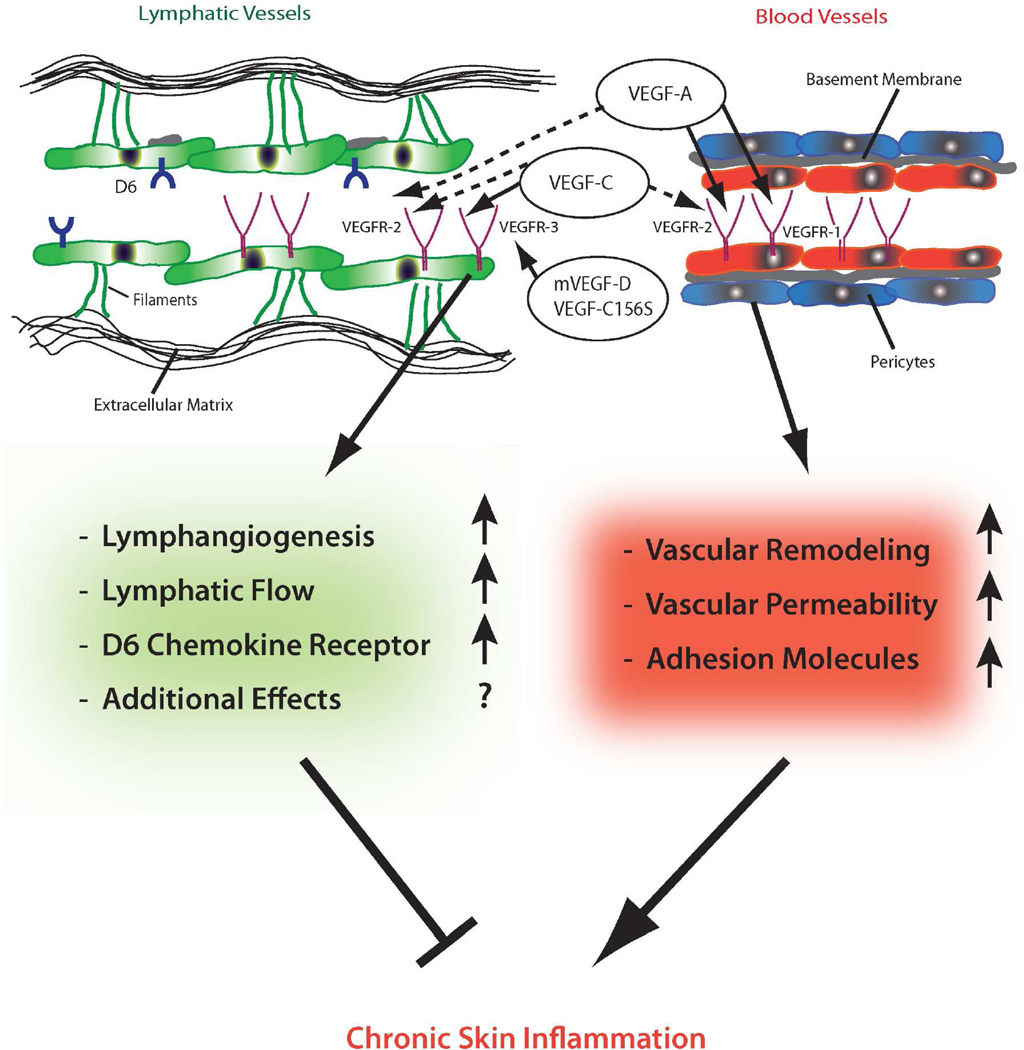
The blood and lymphatic vasculature have an important role in skin homeostasis. Angiogenesis and lymphangiogenesis-the growth of new vessels from existing ones-have received tremendous interest because of their role in promoting cancer spread. However, there is increasing evidence that both vessel types also have a major role in acute and chronic inflammatory disorders. Vessels change their phenotype during inflammation (vascular remodeling). In inflamed skin, vascular remodeling consists of a hyperpermeable, enlarged network of vessels with increased blood flow, and influx of inflammatory cells. During chronic inflammation, the activated endothelium expresses adhesion molecules, cytokines, and other molecules that lead to leukocyte rolling, attachment, and migration into the skin. Recent studies reveal that inhibition of blood vessel activation exerts potent anti-inflammatory properties. Thus, anti-angiogenic drugs might be used to treat inflammatory conditions. In particular, topical application of anti-angiogenic drugs might be ideally suited to circumvent the adverse effects of systemic therapy with angiogenesis inhibitors. Our recent results indicate that stimulation of lymphatic vessel growth and function unexpectedly represents a new approach for treating chronic inflammatory disorders.
Conflict of interest statement
The authors declare no conflict of interest.
Figures
Similar articles
-
Angiogenesis and lymphangiogenesis in inflammatory skin disorders.J Am Acad Dermatol. 2015 Jul;73(1):144-53. doi: 10.1016/j.jaad.2015.03.041. Epub 2015 Apr 25. J Am Acad Dermatol. 2015. PMID: 25922287 Review.
-
Angiogenesis and lymphangiogenesis of skin cancers.Hematol Oncol Clin North Am. 2004 Oct;18(5):1059-70, viii. doi: 10.1016/j.hoc.2004.06.009. Hematol Oncol Clin North Am. 2004. PMID: 15474335 Review.
-
Stimulation of lymphangiogenesis via VEGFR-3 inhibits chronic skin inflammation.J Exp Med. 2010 Sep 27;207(10):2255-69. doi: 10.1084/jem.20100559. Epub 2010 Sep 13. J Exp Med. 2010. PMID: 20837699 Free PMC article.
-
An important role of blood and lymphatic vessels in inflammation and allergy.J Allergy (Cairo). 2013;2013:672381. doi: 10.1155/2013/672381. Epub 2013 Jan 31. J Allergy (Cairo). 2013. PMID: 23431319 Free PMC article.
-
Chapter 1. Inflammation, angiogenesis, and lymphangiogenesis.Methods Enzymol. 2008;445:1-25. doi: 10.1016/S0076-6879(08)03001-2. Methods Enzymol. 2008. PMID: 19022053
Cited by
-
Microvascular imaging and monitoring of hemodynamic changes in the skin during arterial-venous occlusion using multispectral raster-scanning optoacoustic mesoscopy.Photoacoustics. 2021 Apr 20;22:100268. doi: 10.1016/j.pacs.2021.100268. eCollection 2021 Jun. Photoacoustics. 2021. PMID: 34026491 Free PMC article.
-
Skin-on-a-Chip Technology: Microengineering Physiologically Relevant In Vitro Skin Models.Pharmaceutics. 2022 Mar 21;14(3):682. doi: 10.3390/pharmaceutics14030682. Pharmaceutics. 2022. PMID: 35336056 Free PMC article. Review.
-
Blood vessel remodeling in the cerebral cortex induced by binge alcohol intake in mice.Toxicol Res. 2022 Dec 21;39(1):169-177. doi: 10.1007/s43188-022-00164-y. eCollection 2023 Jan. Toxicol Res. 2022. PMID: 36726835 Free PMC article.
-
Interaction Between Blood Vasculatures and Lymphatic Vasculatures During Inflammation.J Inflamm Res. 2023 Aug 4;16:3271-3281. doi: 10.2147/JIR.S414891. eCollection 2023. J Inflamm Res. 2023. PMID: 37560514 Free PMC article. Review.
-
Inflammation and the blood microvascular system.Cold Spring Harb Perspect Biol. 2014 Oct 23;7(1):a016345. doi: 10.1101/cshperspect.a016345. Cold Spring Harb Perspect Biol. 2014. PMID: 25384307 Free PMC article. Review.
References
-
- Abraham DJ, Krieg T, Distler J, Distler O. Overview of pathogenesis of systemic sclerosis. Rheumatology (Oxford) 2009;48(Suppl 3):iii3–iii7. - PubMed
-
- Adams RH, Alitalo K. Molecular regulation of angiogenesis and lymphangiogenesis. Nat Rev Mol Cell Biol. 2007;8:464–478. - PubMed
-
- Agha-Majzoub R, Becker RP, Schraufnagel DE, Chan LS. Angiogenesis: the major abnormality of the keratin-14 IL-4 transgenic mouse model of atopic dermatitis. Microcirculation. 2005;12:455–476. - PubMed
-
- Aird WC. Phenotypic heterogeneity of the endothelium: I. Structure, function, and mechanisms. Circ Res. 2007;100:158–173. - PubMed
Publication types
MeSH terms
Substances
Grants and funding
LinkOut - more resources
Full Text Sources
Other Literature Sources
