. 2011 Dec;15(1):53-62.
doi: 10.1038/jidsymp.2011.6.

Neurovascular and neuroimmune aspects in the pathophysiology of rosacea

Affiliations

Neurovascular and neuroimmune aspects in the pathophysiology of rosacea

Verena D Schwab et al. J Investig Dermatol Symp Proc. 2011 Dec.

Abstract

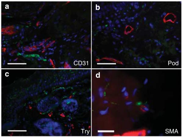
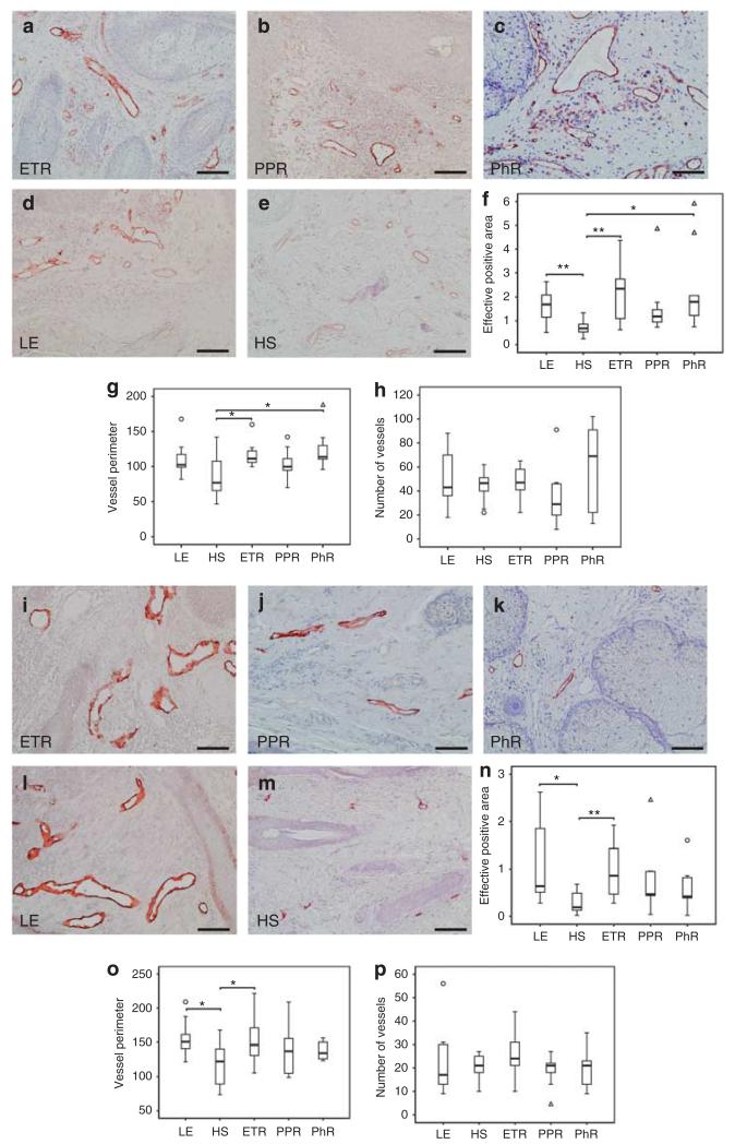
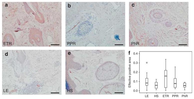
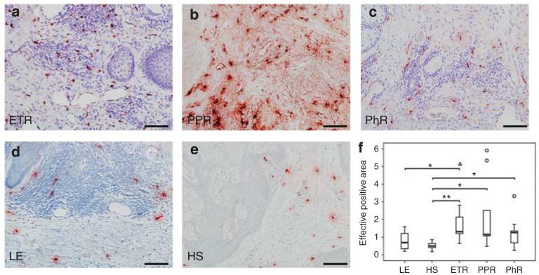
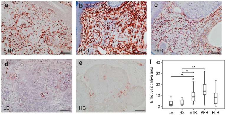
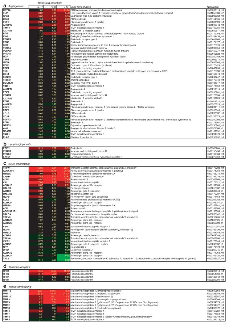
Rosacea is a common skin disease with a high impact on quality of life. Characterized by erythema, edema, burning pain, immune infiltration, and facial skin fibrosis, rosacea has all the characteristics of neurogenic inflammation, a condition induced by sensory nerves via antidromically released neuromediators. To investigate the hypothesis of a central role of neural interactions in the pathophysiology, we analyzed molecular and morphological characteristics in the different subtypes of rosacea by immunohistochemistry, double immunofluorescence, morphometry, real-time PCR, and gene array analysis, and compared the findings with those for lupus erythematosus or healthy skin. Our results showed significantly dilated blood and lymphatic vessels. Signs of angiogenesis were only evident in phymatous rosacea. The number of mast cells and fibroblasts was increased in rosacea, already in subtypes in which fibrosis is not clinically apparent, indicating early activation. Sensory nerves were closely associated with blood vessels and mast cells, and were increased in erythematous rosacea. Gene array studies and qRT-PCR confirmed upregulation of genes involved in vasoregulation and neurogenic inflammation. Thus, dysregulation of mediators and receptors implicated in neurovascular and neuroimmune communication may be crucial at early stages of rosacea. Drugs that function on neurovascular and/or neuroimmune communication may be beneficial for the treatment of rosacea.


Figures

Figure 1
Figure 1. Association of different vascular and immune structures with sensory nerves in human facial skin of rosacea patients, as shown by double immunofluorescence staining
Colocalization of sensory nerves (PGP9.5) was determined in combination with CD31 for blood vessels (a), podoplanin (Pod) for lymphatic vessels (b), tryptase (Try) for mast cells (c), and smooth muscle actin (SMA) for myofibroblasts or blood vessels (d). Our data show a close anatomical association of unmyelinated nerves, especially with blood vessels and mast cells, and less with lymphatic vessels or myofibroblasts (bar = 300 μm; ac and bar = 100 μm; d).
Figure 2
Figure 2. Density of blood and lymphatic vessels in human skin, as shown by immunohistochemistry and quantitative analysis of stained dermis
Immunoreactivity for CD31 and podoplanin was observed in erythematous rosacea (ETR, n = 9), papulopustular rosacea (PPR, n = 9), phymatous rosacea (PhR, n = 9), lupus erythematosus (LE, n = 9), and healthy skin staining (HS, n = 10; bar = 100 μm; ae and im). (f) PhR showed strongest statistically significant augmentation of CD31-positive tissue, followed by ETR (fh; np; *P<0.05; **P<0.01). (g) Vessel perimeter measurement showed significant vasodilation in ETR and PhR, whereas the number of vessels (h) was not increased. A tendency toward angiogenesis was only observed in PhR. Augmentation in lymph vessel surface was statistically significant exclusively in ETR (n). Lymph vessel circumference measurements showed significant vasodilation in ETR (o). Number of lymphatic vessels was not increased in any subtype (p), unfilled triangles and circles represent outliers (fh; np).
Figure 3
Figure 3. Localization and density of myelinated sensory nerves (NF200) in human skin, as shown by immunohistochemistry and quantitative analysis of stained dermis
Immunoreactivity for neurofilament was observed in erythematous rosacea (ETR, n = 9), papulopustular rosacea (PPR; n = 9), phymatous rosacea (PhR; n = 9), lupus erythematosus (LE; n = 9), and healthy skin (HS; n = 10; bar = 100 μm; ae). There was a marked but not statistically significant increase of nerves in ETR (× 2.34) followed by a gradual decrease. Increase of neurofilament-positive nerves was comparable in PPR and LE (unfilled circle represents outlier) (f).
Figure 4
Figure 4. Localization and density of mast cells in human facial skin as shown by immunohistochemistry and quantitative analysis of stained dermis
Immunoreactivity for tryptase was observed in erythematous rosacea (ETR, n = 9), papulopustular rosacea (PPR, n = 9), phymatous rosacea (PhR, n = 9),lupus erythematosus (LE; n = 9), and healthy skin (HS, n = 10; bar = 100 μm; ae). The increase in mast cell density was statistically significant for all subtypes (ETR × 3.98; PPR × 4.41; PhR × 2.54), whereas mast cell density did not increase in LE (*P<0.05, **P<0.01; unfilled circles and triangle represent outliers) (f).
Figure 5
Figure 5. Localization and density of fibroblasts (FBs)/cytes and mesenchymal structures of blood vessels in human skin, as shown by immunohistochemistry and quantitative analysis of stained dermis
Immunoreactivity for vimentin was observed in erythematous rosacea (ETR, n = 9), papulopustular rosacea(PPR, n = 9), phymatous rosacea (PhR, n = 9), lupus erythematosus (LE; n = 9), and healthy skin (HS; n = 10; bar = 100 μm; ae). Density of FBs was increased in all subtypes, but significantly so in ETR (× 2.9) and PPR (× 3.94). Skin of lupus patients showed decreased density of FBs/cytes as compared with healthy human skin (*P<0.05; **P<0.01; unfilled circle represents outlier) (f).
Figure 6
Figure 6. Detection of mRNA levels of genes relevant for neuroimmune and neurovascular interaction as determined by RT-PCR
(a) Fold induction of genes involved in angiogenesis. Modulation of expression was detectable in phymatous rosacea (PhR, n = 6), but not in erythematous rosacea (ETR, n = 11) and papulopustular rosacea (PPR, n = 11). (b) Fold induction of genes involved in lymphangiogenesis. RT-PCR showed upregulation of podoplanin, but downregulation of LYVE1. (c) Fold induction of genes involved in neurovascular interaction. RT-PCR showed marked modulation of gene expression of different neuropeptides and their corresponding receptors in all subtypes. (d) Fold induction of histamine receptors. HRH3 was upregulated. (e) Fold induction of genes involved in tissue remodeling. RT-PCR shows evidence of a strong enhancement of matrix metalloproteinases (MMPs), whereas their inhibitors are downregulated.

References

    1. Amălinei C, Căruntu ID, Giuşcă SE, et al. Matrix metalloproteinases involvement in pathologic conditions. Rom J Morphol Embryol. 2010;51:215–28. - PubMed
    1. Amerini S, Ziche M, Greiner ST, et al. Effects of substance P on mesenteric lymphatic contractility in the rat. Lymphat Res Biol. 2004;2:2–10. - PubMed
    1. Arck PC, Slominski A, Theoharides TC, et al. Neuroimmunology of stress: skin takes center stage. J Invest Dermatol. 2006;126:1697–704. - PMC - PubMed
    1. Aroni K, Tsagroni E, Kavantzas N, et al. A study of the pathogenesis of rosacea: how angiogenesis and mast cells may participate in a complex multifactorial process. Arch Dermatol Res. 2008;300:125–31. - PubMed
    1. Aroni K, Tsagroni E, Lazaris AC, et al. Rosacea: a clinicopathological approach. Dermatology. 2004;209:177–82. - PubMed

MeSH terms