. 2011 Dec;3(12):1224-32.
doi: 10.1039/c1ib00064k. Epub 2011 Nov 10.

Magnetic manipulation and spatial patterning of multi-cellular stem cell aggregates

Affiliations

Magnetic manipulation and spatial patterning of multi-cellular stem cell aggregates

Andrés M Bratt-Leal et al. Integr Biol (Camb). 2011 Dec.

Abstract

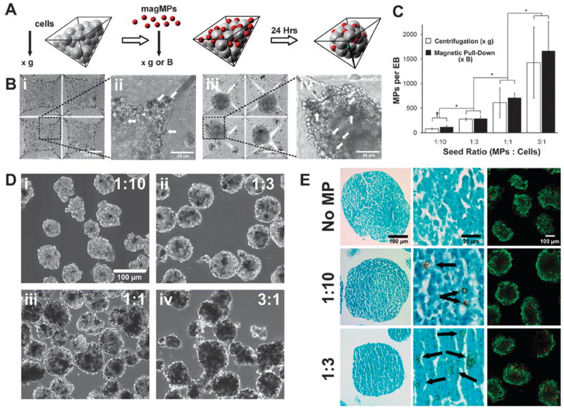
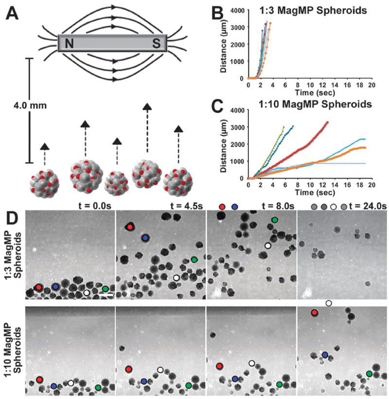
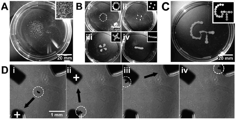
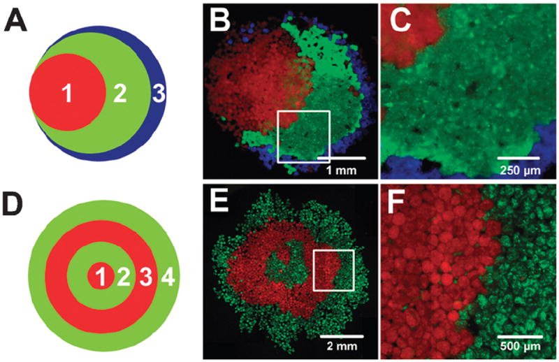
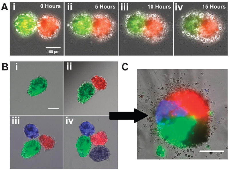
The controlled assembly and organization of multi-cellular systems to mimic complex tissue structures is critical to the engineering of tissues for therapeutic and diagnostic applications. Recent advances in micro-scale technologies to control multi-cellular aggregate formation typically require chemical modification of the interface between cells and materials and lack multi-scale flexibility. Here we demonstrate that simple physical entrapment of magnetic microparticles within the extracellular space of stem cells spheroids during initial formation enables scaffold-free immobilization, translocation and directed assembly of multi-cellular aggregates across multiple length and time scales, even under dynamic suspension culture conditions. The response of aggregates to externally applied magnetic fields was a direct function of microparticle incorporation, allowing for rapid and transient control of the extracellular environment as well as separation of heterogeneous populations. In addition, spatial patterning of heterogeneous spheroid populations as well as individual multi-cellular aggregates was readily achieved by imposing temporary magnetic fields. Overall, this approach provides novel routes to examine stem cell differentiation and tissue morphogenesis with applications that encompass the creation of new model systems for developmental biology, scaffold-free tissue engineering strategies and scalable bioprocessing technologies.


Figures

Fig. 1
Fig. 1
Magnetic MPs are incorporated in a dose dependent manner. (a) MagMPs were incorporated within stem cell spheroids within PDMS microwells (b) MagMPs (arrows) were observed after ESC and magMP centrifugation (i,ii) as well as throughout the spheroids that were formed after 18–24 h of culture (iii,iv). Scale bars, 200 μm (i,iv) and 50 μm (ii,iii) (c) Spheroids were formed using centrifugation or magnetic pull-down and the number of MPs incorporated per spheroid was determined after 24 h of culture. *p ≤ 0.05 Comparison of loading ratios, †p ≤ 0.05 comparison between pull-down and centrifugation. (d) EBs formed at cell seeding ratios of 1 : 10 (i), 1 : 3 (ii), 1 : 1 (iii), 3 : 1 (iv) were of similar shape and size and dark regions of magMPs increased with the seeding ratio. Scale bar 100μm. (e) Histological sections stained with Fast Green stain revealed that magMP incorporation did not disrupt cellular arrangement or morphology. No differences in cell viability were observed for any of the groups (LIVE/DEAD assay, dead cells labeled red and live cells labeled green). Scale bars 100 μm (first and third columns) and 20 μm (middle column).
Fig. 2
Fig. 2
Spheroids with magMPs respond in a dose dependent fashion to magnetic fields. (a) Spheroids can be directed to move in a dish by an externally applied magnetic field. (b,c) Spheroids formed with a higher ratio (1 : 3) of magMPs to cells move faster and more uniformly relative to spheroids formed with a 1 : 10 seed ratio. (d) The distance moved between frames was tracked and suggests that the populations could be sorted based on magnetic sensitivity alone. Scale bar 500 μm.
Fig. 3
Fig. 3
Spheroid location can be controlled in dynamic suspension culture. (a) Without magnets, spheroids cultured on a rotary orbital shaker (45 RPM) cluster in the center of the dish. Scale bar 20 mm. (b) The placement magnets on the lid of the dish (configuration shown in insets) could be used to confine the spheroids to (i) a defined radius, (ii) distinct islands, (iii) a four-leaf clover, (iv) or a line configuration. Scale bar 20 mm. (c). More complicated patterns, including the Georgia Tech logo, could be made at slower culture speeds. Scale bar 20 mm. (d) In addition, the location of a single spheroid could be manipulated back and forth between iron pillars embedded in PDMS. The iron pillars were alternately magnetized (demonstrated by a “+” label), resulting in attraction of the spheroid to the magnetized pillar (i–iv). Scale bar 1 mm.
Fig. 4
Fig. 4
Large populations of magMP spheroids were manipulated to form macroscopic patterns including a “stacked Venn diagram” shape (a–c) or a “bullseye” pattern (d–f). Populations of CellTracker labeled EBs were added sequentially as shown in panels a and d and were aggregated using a magnet applied underneath the Petri dish. Mixing between the layers was limited as demonstrated in panel c and f, magnified from the boxed areas in b and e. Scale bars 1mm (b), 250 μm (c), 2 mm (e), 500 μm (f).
Fig. 5
Fig. 5
Merging of EBs can be spatially controlled on the single aggregate level. (a) The merging process of two spheroids was visualized over a period of 15 h using time lapse microscopy. Fluorescently labeled stem cell spheroids were brought in contact with one and other using a magnetic probe (i) and began to merge after 5 h of culture (ii). The spheroids continued to merge after 10 h (iii) and became more rounded after 15 h (iv). Scale bar 100 μm. (b) Merging of more than two spheroids could also be controlled by magnetic manipulation. Spheroids were drawn together magnetically in a sequential manner (i–iv) which maintained the position of the spheroids relative to each other. Scale bar 100 μm. (c) After 24 h of culture 4 spheroids merged to create a single construct consisting of four distinct quadrants of fluorescence. Scale bar 100 μm.

References

    1. Gumbiner BM. Cell adhesion: the molecular basis of tissue architecture and morphogenesis. Cell. 1996;84:345–57. - PubMed
    1. Khetani SR, Bhatia SN. Microscale culture of human liver cells for drug development. Nat Biotechnol. 2008;26:120–6. - PubMed
    1. Langer R, Vacanti JP. Tissue engineering. Science. 1993;260:920–6. - PubMed
    1. Takeichi M. Cadherins: a molecular family important in selective cell-cell adhesion. Annu Rev Biochem. 1990;59:237–52. - PubMed
    1. Hynes RO, Lander AD. Contact and adhesive specificities in the associations, migrations, and targeting of cells and axons. Cell. 1992;68:303–22. - PubMed

Publication types

MeSH terms