Clinical Trial
. 2011 Nov 22;77(21):1877-86.
doi: 10.1212/WNL.0b013e318239f7ef. Epub 2011 Nov 9.

Intrathecal effects of daclizumab treatment of multiple sclerosis

Affiliations
Clinical Trial

Intrathecal effects of daclizumab treatment of multiple sclerosis

B Bielekova et al. Neurology. .

Abstract

Objectives: We previously reported that daclizumab, a humanized monoclonal antibody against CD25, reduced contrast-enhancing lesions (CEL) in patients with multiple sclerosis (MS) who were suboptimal responders to interferon-β and that this response correlated with expansion of CD56(bright) NK cells. These data have been reproduced in a placebo-controlled multicenter trial (CHOICE study). The current study investigates whether daclizumab monotherapy reduces CEL in untreated patients with relapsing-remitting MS (RRMS) and the effects of daclizumab on the intrathecal immune system.

Methods: Sixteen patients with RRMS with high inflammatory activity were enrolled in an open-label, baseline-vs-treatment, phase II trial of daclizumab monotherapy for 54 weeks and followed by serial clinical and MRI examinations and immunologic biomarkers measured in the whole blood and CSF.

Results: The trial achieved predefined outcomes. There was an 87.7% reduction in brain CEL (primary) and improvements in Multiple Sclerosis Functional Composite (secondary), Scripps Neurologic Rating Scale, and Expanded Disability Status Scale (tertiary) outcomes. There was significant expansion of CD56(bright) NK cells in peripheral blood and CSF, with resultant decrease in T cells/NK cells and B cells/NK cells ratios and IL-12p40 in the CSF. Surprisingly, CD25 Tac epitope was equally blocked on the immune cells in the CSF and in peripheral blood.

Conclusions: Daclizumab monotherapy inhibits formation of MS plaques in patients with RRMS and immunoregulatory NK cells may suppress activation of pathogenic immune responses directly in the CNS compartment.

Classification of evidence: The study provides Class III evidence that daclizumab reduces the number of contrast-enhancing lesions in treatment-naive patients with RRMS over a 54-week period.


Figures

Figure 1
Figure 1. Trial design and flow diagram
(A) Trial design: patients had 4 monthly clinical and MRI examinations during untreated “baseline” before daclizumab dosing was initiated at 1 mg/kg IV. The first 2 doses were 2 weeks apart, after which patients received daclizumab infusion every 4 weeks for total of 54 weeks. Although clinical and MRI examinations were performed monthly, only 3 predefined 12-week periods were considered in data analysis: the baseline period (week −12 through week 0), primary treatment phase (weeks 18–30), and completion of treatment phase (weeks 42–54). For all outcomes, patient-specific averages of 4 MRI/clinical examinations were computed for all 3 phases and analyzed as described in Methods. For immunologic outcomes, matched CSF and blood samples were collected once during pretreatment baseline and once after week 26 of treatment. There was optional CSF collection at patient's discretion at week 6 of daclizumab treatment. (B) CONSORT statement 2010 flow diagram.
Figure 2
Figure 2. MRI and clinical outcomes
(A–D) Period averages were computed for each patient as described in Methods and are depicted as dot and line plots. Group data are depicted as open box blots, with group medians represented by black and means by red horizontal lines. Statistically significant differences are highlighted by asterisk (*p ≤ 0.01 and **p ≤ 0.001). CEL = contrast-enhancing lesion; EDSS = Expanded Disability Status Scale; MSFC = Multiple Sclerosis Functional Composite; NRS = Neurological Rating Scale.
Figure 3
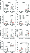
Figure 3. Effect of daclizumab therapy on cellular subpopulations
(A–H) Patient-specific data are depicted as dot and line blots. Group data are depicted as open box blots, with group medians represented by black and means by red horizontal lines. Statistically significant differences are highlighted with exact p values depicted in the figure. MFI= mean fluorescent intensity.
Figure 4
Figure 4. Representative example of raw flow cytometry data
Flow cytometry staining on immune cells (CD4+ T cells: first 2 rows, CD3+ T cells: third row, and CD3−/CD56+ NK cells: fourth row) in the blood (left panels) and paired CSF samples (right panels) isolated during pretreatment baseline and after week 26 of daclizumab therapy in a representative patient. The red arrow in the first row highlights higher expression of CD122 (interleukin [IL] -2R-β chain) in the CSF as compared to blood. In the second row, T cells positive for CD25 (both Tac and 7G7 epitopes) are highlighted in red. In the third row, the red arrow highlights significantly higher expression of CXCR3 and CCR5 chemokine receptors on the T cells in the CSF as compared to blood, but there is no change with daclizumab therapy. Quadrant gates were set based on appropriate isotype controls for each staining combination. In the fourth row, CD56bright NK cells (highlighted in red) are gated in the blood based on their higher expression of CD56 and CD44 in comparison to CD56dim NK cell counterparts. However, in the CSF, all NK cells express uniformly high CD44. The proportion of NK cells that were gated as CD56bright both in the blood and CSF are depicted as number in each contour blot.
Figure 5
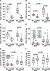
Figure 5. Effect of daclizumab therapy on interleukin (IL)-2R chains
(A–F) Patient-specific data are depicted as dot and line blots. Group data are depicted as open box blots, with group medians represented by black and means by red horizontal lines. Statistically significant differences are highlighted with exact p values depicted in the figure. MFI= mean fluorescent intensity.

References

    1. Bielekova B, Richert N, Howard T, et al. Humanized anti-CD25 (daclizumab) inhibits disease activity in multiple sclerosis patients failing to respond to interferon-beta. Proc Natl Acad Sci USA 2004;101:8705–8708 - PMC - PubMed
    1. Bielekova B, Howard T, Packer AN, et al. Effect of anti-CD25 antibody daclizumab in the inhibition of inflammation and stabilization of disease progression in multiple sclerosis. Arch Neurol 2009;66:483–489 - PMC - PubMed
    1. Rose JW, Burns JB, Bjorklund J, Klein J, Watt HE, Carlson NG. Daclizumab phase II trial in relapsing and remitting multiple sclerosis: MRI and clinical results. Neurology 2007;69:785–789 - PubMed
    1. Bielekova B, Catalfamo M, Reichert-Scrivner S, et al. Regulatory CD56bright natural killer cells mediate immunomodulatory effects of IL-2R-alpha-targeted therapy (daclizumab) in multiple sclerosis. PNAS 2006;103:5941–5946 - PMC - PubMed
    1. Wynn D, Kaufman M, Montalban X, et al. Daclizumab in active relapsing multiple sclerosis (CHOICE study): a phase 2, randomised, double-blind, placebo-controlled, add-on trial with interferon beta. Lancet Neurol 2010;9:381–390 - PubMed

Publication types

MeSH terms