

Identification of two new arthritis severity loci that regulate levels of autoantibodies, interleukin-1β, and joint damage in pristane- and collagen-induced arthritis
- PMID: 22076633
- PMCID: PMC3288617
- DOI: 10.1002/art.33468
Identification of two new arthritis severity loci that regulate levels of autoantibodies, interleukin-1β, and joint damage in pristane- and collagen-induced arthritis
Abstract
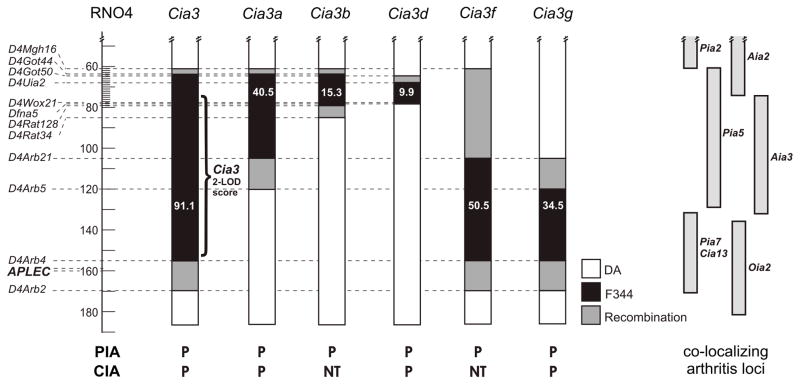
Objective: Cia3 is a locus on rat chromosome 4 that regulates severity and joint damage in collagen- and pristane-induced arthritis (CIA and PIA). This study was undertaken to refine the Cia3 gene-containing interval toward gene identification and obtain insights into its mode of action.
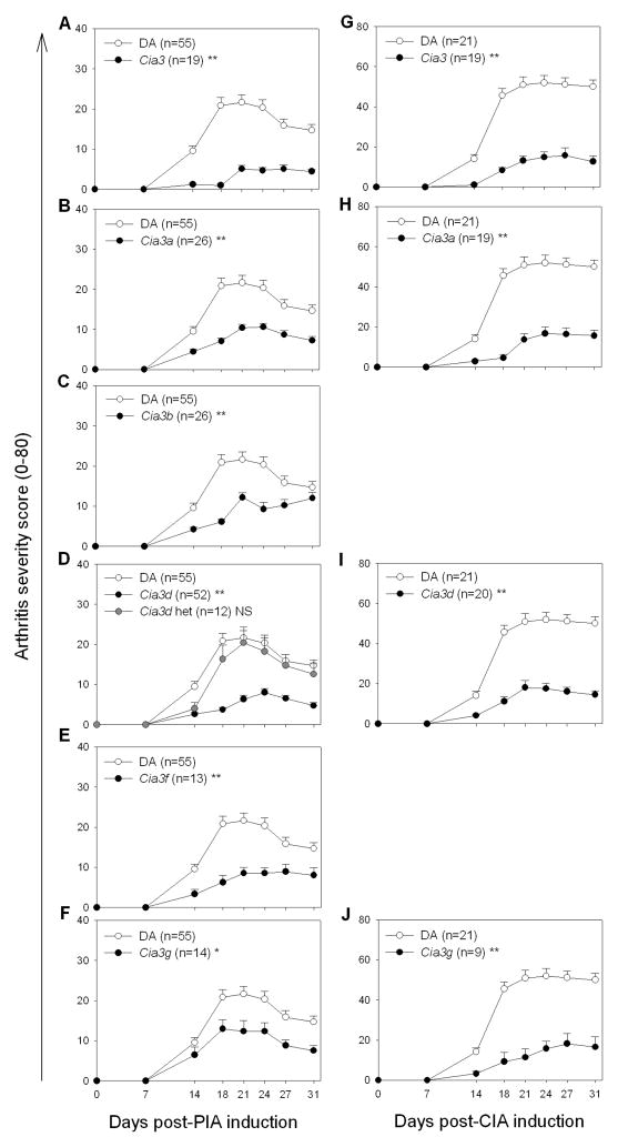
Methods: Five DA.F344(Cia3) subcongenic rat strains were generated and studied using the PIA and CIA models. Levels of antibodies against type II collagen (both allo- and autoantibodies) were measured. Joints and synovial tissue were collected 32 days after the induction of PIA (chronic stage) for histologic and quantitative polymerase chain reaction analysis of interleukin-1β (IL-1β) and matrix metalloproteinase (MMP) levels.
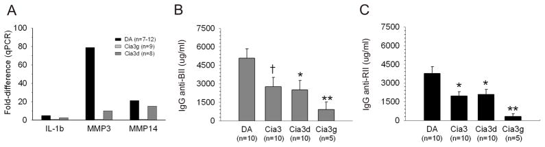
Results: Three subcongenic strains sharing the centromeric Cia3d interval were protected and 2 subcongenic strains sharing the telomeric Cia3g interval, which did not overlap with Cia3d, were also protected, developing significantly less severe CIA and PIA. Normal joint architecture was preserved in DA.F344(Cia3) and DA.F344(Cia3d) congenic rats with PIA, while DA rats had pronounced synovial hyperplasia, angiogenesis, inflammatory infiltration, and bone or cartilage erosions. The DA.F344(Cia3d) and DA.F344(Cia3g) strains had significantly lower synovial levels of IL-1β (5-fold and nearly 2-fold, respectively [the latter not reaching statistical significance]), MMP-1 (expressed predominantly in DA rats), MMP-3 (79-fold and 8-fold, respectively), and MMP-14 (21-fold and 1.4-fold, respectively) and reduced levels of pathogenic autoantibodies against type II collagen, compared with DA rats.
Conclusion: We have identified 2 new arthritis severity and articular damage loci within Cia3. These loci regulate pathogenic processes in 2 different models of rheumatoid arthritis, and the identification of these genes has the potential to generate new targets for therapies aimed at reducing disease severity and articular damage, and may additionally have prognostic value.
Copyright © 2012 by the American College of Rheumatology.
Figures

Similar articles
-
The non-MHC quantitative trait locus Cia5 contains three major arthritis genes that differentially regulate disease severity, pannus formation, and joint damage in collagen- and pristane-induced arthritis.J Immunol. 2005 Jun 15;174(12):7894-903. doi: 10.4049/jimmunol.174.12.7894. J Immunol. 2005. PMID: 15944295
-
The non-major histocompatibility complex quantitative trait locus Cia10 contains a major arthritis gene and regulates disease severity, pannus formation, and joint damage.Arthritis Rheum. 2005 Jan;52(1):322-32. doi: 10.1002/art.20782. Arthritis Rheum. 2005. PMID: 15641042
-
Cia25 on rat chromosome 12 regulates severity of autoimmune arthritis induced with pristane and with collagen.Ann Rheum Dis. 2007 Jul;66(7):952-7. doi: 10.1136/ard.2006.066225. Epub 2007 Feb 28. Ann Rheum Dis. 2007. PMID: 17329308 Free PMC article.
-
Arthritis severity locus Cia4 is an early regulator of IL-6, IL-1β, and NF-κB activators' expression in pristane-induced arthritis.Physiol Genomics. 2013 Jul 2;45(13):552-64. doi: 10.1152/physiolgenomics.00029.2013. Epub 2013 May 21. Physiol Genomics. 2013. PMID: 23695883 Free PMC article.
-
Modulation of multiple experimental arthritis models by collagen-induced arthritis quantitative trait loci isolated in congenic rat lines: different effects of non-major histocompatibility complex quantitative trait loci in males and females.Arthritis Rheum. 2002 Aug;46(8):2225-34. doi: 10.1002/art.10439. Arthritis Rheum. 2002. PMID: 12209529
Cited by
-
Whole-genome sequences of DA and F344 rats with different susceptibilities to arthritis, autoimmunity, inflammation and cancer.Genetics. 2013 Aug;194(4):1017-28. doi: 10.1534/genetics.113.153049. Epub 2013 May 20. Genetics. 2013. PMID: 23695301 Free PMC article.
-
Analyses of synovial tissues from arthritic and protected congenic rat strains reveal a new core set of genes associated with disease severity.Physiol Genomics. 2013 Nov 15;45(22):1109-22. doi: 10.1152/physiolgenomics.00108.2013. Epub 2013 Sep 17. Physiol Genomics. 2013. PMID: 24046282 Free PMC article.
-
Resolvin D1 suppresses pannus formation via decreasing connective tissue growth factor caused by upregulation of miRNA-146a-5p in rheumatoid arthritis.Arthritis Res Ther. 2020 Mar 27;22(1):61. doi: 10.1186/s13075-020-2133-2. Arthritis Res Ther. 2020. PMID: 32216830 Free PMC article.
-
Liver X receptor regulates rheumatoid arthritis fibroblast-like synoviocyte invasiveness, matrix metalloproteinase 2 activation, interleukin-6 and CXCL10.Mol Med. 2012 Sep 7;18(1):1009-17. doi: 10.2119/molmed.2012.00173. Mol Med. 2012. PMID: 22634718 Free PMC article.
-
Effects by periodontitis on pristane-induced arthritis in rats.J Transl Med. 2016 Nov 3;14(1):311. doi: 10.1186/s12967-016-1067-6. J Transl Med. 2016. PMID: 27809921 Free PMC article.
References
-
- Gregersen PK, Plenge RM, Gulko PS. Genetics of rheumatoid arthritis. In: Firestein G, Panayi G, Wollheim FA, editors. Rheumatoid arthritis. 2. New York: Oxford University Press; 2006. pp. 3–14.
-
- Marinou I, Maxwell JR, Wilson AG. Genetic influences modulating the radiological severity of rheumatoid arthritis. Ann Rheum Dis. 2010;69(3):476–82. - PubMed
-
- Gulko PS, Kawahito Y, Remmers EF, Reese VR, Wang J, Dracheva SV, et al. Identification of a new non-major histocompatibility complex genetic locus on chromosome 2 that controls disease severity in collagen- induced arthritis in rats. Arthrititis Rheum. 1998;41(12):2122–31. - PubMed
Publication types
MeSH terms
Substances
Grants and funding
LinkOut - more resources
Full Text Sources
Miscellaneous
