. 2012 Jan 25;53(1):361-73.
doi: 10.1167/iovs.11-8635.

Prominin-1 localizes to the open rims of outer segment lamellae in Xenopus laevis rod and cone photoreceptors

Affiliations

Prominin-1 localizes to the open rims of outer segment lamellae in Xenopus laevis rod and cone photoreceptors

Zhou Han et al. Invest Ophthalmol Vis Sci. .

Abstract

Purpose: Prominin-1 expresses in rod and cone photoreceptors. Mutations in the prominin-1 gene cause retinal degeneration in humans. In this study, the authors investigated the expression and subcellular localization of xlProminin-1 protein, the Xenopus laevis ortholog of prominin-1, in rod and cone photoreceptors of this frog.

Methods: Antibodies specific for xlProminin-1 were generated. Immunoblotting was used to study the expression and posttranslational processing of xlProminin-1 protein. Immunocytochemical light and electron microscopy and transgenesis were used to study the subcellular distribution of xlProminin-1.

Results: xlProminin-1 is expressed and is subject to posttranslational proteolytic processing in the retina, brain, and kidney. xlProminin-1 is differently expressed and localized in outer segments of rod and cone photoreceptors of X. laevis. Antibodies specific for the N or C termini of xlProminin-1 labeled the open rims of lamellae of cone outer segments (COS) and the open lamellae at the base of rod outer segments (ROS). By contrast, anti-peripherin-2/rds antibody, Xper5A11, labeled the closed rims of cone lamellae adjacent to the ciliary axoneme and the rims of the closed ROS disks. The extent of labeling of the basal ROS by anti-xlProminin-1 antibodies varied with the light cycle in this frog. The entire ROS was also faintly labeled by both antibodies, a result that contrasts with the current notion that prominin-1 localizes only to the basal ROS.

Conclusions: These findings suggest that xlProminin-1 may serve as an anti-fusogenic factor in the regulation of disk morphogenesis and may help to maintain the open lamellar structure of basal ROS and COS disks in X. laevis photoreceptors.


Figures

Figure 1.
Figure 1.
Detection of xlProminin-1 in membrane fractions of X. laevis retina, brain, and kidney by immunoblotting. Membrane proteins extracted from X. laevis retina OS, IF, and from brain and kidney were immunoblotted with either anti–xlProminin-1 N terminus antibody αPN or anti–xlProminin-1 C terminus antibody αPC for the presence of xlProminin-1 protein. A product of approximately 95 kDa was detected in OS and IF fractions of the retina by both αPN and αPC antibodies (filled arrows). PNGase F treatment reduced the molecular weight of this protein to approximately 80 kDa. This product may represent full-length xlProminin-1. No 95-kDa product is detected with either αPN or αPC in membrane proteins extracted from the brain or kidney. A minor band (approximately 45 kDa, hollow arrow) was detected by αPN antibody in the IF fraction of the retina, and the membrane protein from brain and kidney. The molecular weight of this protein was reduced to approximately 30 kDa after PNGase F treatment. A low molecular band (asterisk) detected by both αPN and αPC in the IF fraction of retina may represent the degradation products of xlProminin-1 because this band contains epitopes from both N and C termini of xlProminin-1, but with a much lower molecular weight (approximately 12 kDa) than that of the full-length protein. Anti–acetylated α-tubulin antibody was used as a loading control.
Figure 2.
Figure 2.
Double immunolabeling of X. laevis photoreceptors with anti–xlProminin-1 N terminus antibody αPN and anti–α-tubulin antibody. Sections of X. laevis retina were double immunolabeled with αPN and anti–acetylated α-tubulin antibody. (A) The COS is brightly labeled with αPN (green) predominantly on one side (arrows). The base of the ROS is also faintly labeled with αPN as a thin band (arrowheads). Distal ROS is diffusively labeled with αPN at low intensity. The labeling intensity of αPN on COS is much greater than on ROS. (B) The same section of retina was labeled with anti–acetylated α-tubulin antibody (red). Axonemes of the connecting cilia of both rods (arrowheads) and cones (arrows) are labeled with that antibody. (C) A longitudinally sectioned cone cell on the upper part of the image shows clearly separated labeling of xlProminin-1 and α-tubulin. Labeling of αPN on the rims is confined to one side of the COS (arrowhead). A differently oriented cell is also seen on the lower left. (D) Nomarski view of the same retina section to show the morphology of cells. (E) Superimposed image to show the relative position of αPN and anti–α-tubulin immunolabeling. αPN labels the lamellar rims opposite the axoneme of the COS. (F) Cross-section of a tadpole retina labeled with αPN (green) and Texas Red-X conjugated wheat germ agglutinin (red) that binds to glycosylated photopigments. The semicircular pattern of αPN labeling is readily seen in the cross-sectioned COS (arrows), further demonstrating the asymmetrical distribution of xlProminin-1 in this organelle. Scale bars: 2 μm (A, B, D, E); 5 μm (C, F).
Figure 3.
Figure 3.
Double immunolabeling of X. laevis photoreceptors with anti–xlProminin-1 C terminus antibody αPC and anti–α-tubulin antibody. (A) The COS is brightly labeled with αPC (green) predominantly on one side. The base of a ROS (arrowheads) is also labeled with αPC as a thin faint band. Distal ROS is diffusively labeled by αPC, as it is by αPN (Fig. 2). (B) Enlarged, brightened, and contrast-enhanced images of boxed areas in (A) to show the basal labeling of ROS (arrowheads). (C) The same retina section was labeled with anti–acetylated α-tubulin antibody (red). Axonemes of the connecting cilia of both rods (arrowheads) and cones (arrows) are labeled. The antibody also labels the filaments that extend from the calycal processes into the inner segment to the level of the external limiting membrane. (D) Nomarski view of the same retina section to show the morphology of cells. (E) Nomarski view of the same enlarged area as in (B). Contours of individual photoreceptors are marked with dashed lined. (F) Superimposed image to show the relative position of αPC and anti–α tubulin immunolabeling. αPC labels the lamellar rims opposite to the axoneme of the COS. CIS, cone inner segment; RIS, rod inner segment. Scale bar, 5 μm.
Figure 4.
Figure 4.
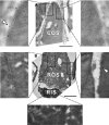
Double immunolabeling of X. laevis photoreceptors with anti–xlProminin-1 N terminus antibody αPN and anti–peripherin-2/rds antibody Xper5A11. (A) The COS is brightly labeled asymmetrically with αPN (green). The base of the ROS is labeled with αPN as a thin, faint band (arrowheads). (B) Enlarged, brightened, and contrast-enhanced images of boxed areas in (A) to show the basal labeling of ROS (arrowheads). (C) The same retina section was labeled with anti–peripherin2/rds antibody Xper5A11 (red). Both COS and ROS are labeled with Xper5A11, but the COS labeling is confined to a thin area along the length of one side of the COS, whereas the ROS are labeled circumferentially except for the interiors of the incisures, which are viewed as parallel unlabeled longitudinal lines. (D) Nomarski image of the same retina section to show the morphology of cells. (E) Nomarski view of the same enlarged area as in (B). Contours of individual photoreceptors are marked with dashed lines. (F) Superimposed image to show the relative position of αPN and Xper5A11 immunolabeling. αPN labels the lamellar rims opposite the side that is labeled with Xper5A11 on the COS. The thin band labeled with αPN at the base of ROS does not overlap with the Xper5A11 labeling of the mature disks above. Scale bar, 5 μm.
Figure 5.
Figure 5.
Double immunolabeling of X. laevis photoreceptors with anti–xlProminin-1 C terminus antibody αPC and anti–peripherin-2/rds antibody Xper5A11. (A) COS is brightly labeled asymmetrically with αPC (green). The base of the ROS is labeled with αPC as a thin faint band (arrowhead). (B) The same retina section was labeled with anti–peripherin2/rds Xper5A11 (red). Both COS and ROS are labeled with Xper5A11, but the COS labeling is confined to a thin area along the length of one side of the COS, whereas the ROS are labeled circumferentially. Unlabeled longitudinal lines in the ROS are the interiors of the incisures. (C) Nomarski view of the same retina section to show the morphology of cells. (D) Superimposed image to show the relative position of αPC and Xper5A11 immunolabeling. αPC labels the lamellar rims opposite the side that is labeled with Xper5A11 on the COS. αPC labeling at the base of ROS does not overlap with Xper5A11 labeling (arrowhead). Scale bars, 5 μm.
Figure 6.
Figure 6.
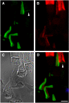
Variation in immunolabeling of X. laevis rods with anti–xlProminin-1 N terminus antibody αPN. (A) Immunolabeling of a retina from a tadpole euthanized at 4 hours before light onset with αPN (green). No labeling is seen at the bases of the ROS. The boxed area is enlarged as shown in (B). (C) Immunolabeling of a retina from a tadpole euthanized at 8 hours after light onset with αPN. Bases of the ROS are clearly labeled (arrowheads). The boxed area is enlarged as shown in (D). Nuclei are labeled with Hoechst 33342 dye (blue). Scale bars, 5 μm.
Figure 7.
Figure 7.
Localization of xlProminin-1 with anti–xlProminin-1 N terminus antibody αPN in X. laevis photoreceptors examined by immunoelectron microscopy of frozen sucrose-embedded retinas. (A) The rims of the disk lamellae of a COS opposite the side of the connecting cilium are labeled with αPN, detected with protein A 6 nm gold (arrowhead). Rims of the disk lamellae adjacent to the connecting cilium are not labeled. (B) Cross-section of a COS. The entire rim except the right side is labeled with αPN, whereas the interior lamellar membranes are unlabeled. (C) Longitudinal section of a rod. The whole ROS is diffusively labeled with αPN at low density. The RIS is only sparsely labeled. Labeling density at the base of this ROS is not higher than at the more distal portions. Boxed areas are enlarged fourfold to show details of representative immunolabeling of the sections. The large holes are artifacts caused by the instability of the fragile sections. C, connecting cilium. Scale bars, 1 μm.
Figure 8.
Figure 8.
Localization of xlProminin-1 with anti–xlProminin-1 C terminus antibody αPC in X. laevis photoreceptors examined by immuno-electron microscopy of frozen sucrose embedded retinas. (A) The rims of the disk lamellae of a COS opposite the side of the connecting cilium are labeled with αPC, detected with protein A 6 nm gold (arrowhead). Rims of the disk lamellae on the same side of the connecting cilium are not labeled. (B) Longitudinal section of a rod. ROS and RIS are not labeled with αPC, in contrast to an adjacent labeled COS membrane (arrowhead). Boxed areas are enlarged fourfold to show the details of typical immunolabeling of the sections. C, connecting cilium. Scale bars, 1 μm.
Figure 9.
Figure 9.
(AD) xlProminin-1-hrGFP II expressed in transgenic X. laevis cones. Expression of the transgene is controlled by a newly isolated promoter XtCAP 1.9 for the X. tropicalis cone arrestin gene (see Supplementary Materials, http://www.iovs.org/lookup/suppl/doi:10.1167/iovs.11-8635/-/DCSupplemental). (A) The hrGFP II-tagged fusion protein localizes primarily to one side of COS (arrowhead). (B) The same section colabeled with anti–peripherin-2/rds antibody Xper5A11 (red). COS was labeled with the antibody and the labeling is confined to a thin area along one side of the COS (arrow). Longitudinal stripes in the adjacent rods result from the peripherin-2/rds localization along the aligned incisures of ROS disks. (C) Nomarski view of the same retina. (D) Superimposed image shows that xlProminin-1-hrGFP II resides on the side of the disk rims opposite peripherin-2/rds. Thus the transgenic hrGFP II-tagged protein expressed in cones distributes to the same sites as the endogenous protein we observed with anti–xlProminin-1 antibodies αPN and αPC. Nuclei are labeled with Hoechst 33342 dye (blue). (EH) xlProminin-1-hrGFP II expressed in transgenic X. laevis rods. Expression of the transgene is controlled by a promoter XOP 1.3 for the X. laevis opsin gene. (E, F) The fusion protein is seen on the plasma membrane of both ROS and RIS (green) and occasionally is observed within the middle of the OS. Some fluorescence is seen within the RIS, presumably representing the trafficking of the newly synthesized protein. (G, H) The fusion protein is occasionally observed at the base of the ROS (arrowhead). Sections were counterstained with Texas Red-X conjugated wheat germ agglutinin (red) to visualize the ROS and post-Golgi membranes. Scale bars, 5 μm.
Figure 10.
Figure 10.
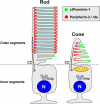
A model of differential distribution of xlProminin-1 and peripherin-2/rds in rod and cone photoreceptors. xlProminin-1 (green) distributes to the outer rims of open disk lamellae of COS and basal disks of ROS. Peripherin-2/rds, in contrast, distributes to the inner rim on the same side as the connecting cilium in COS and also to the entire rim of closed mature disks in ROS. Prominin-1 is also sparsely present throughout the entire disks of ROS. CC, connecting cilium; OD, oil droplet; N, nucleus.

Similar articles

Cited by

References

    1. Bowmaker JK. Visual pigments and colour vision in man and monkeys. J R Soc Med. 1981;74:348–356 - PMC - PubMed
    1. Young RW. Passage of newly formed protein through the connecting cilium of retina rods in the frog. J Ultrastruct Res. 1968;23:462–473 - PubMed
    1. Papermaster DS, Schneider BG, Besharse JC. Vesicular transport of newly synthesized opsin from the Golgi apparatus toward the rod outer segment: ultrastructural immunocytochemical and autoradiographic evidence in Xenopus retinas. Invest Ophthalmol Vis Sci. 1985;26:1386–1404 - PubMed
    1. Roepman R, Wolfrum U. Protein networks and complexes in photoreceptor cilia. Subcell Biochem. 2007;43:209–235 - PubMed
    1. Yau KW. Phototransduction mechanism in retinal rods and cones: the Friedenwald Lecture. Invest Ophthalmol Vis Sci. 1994;35:9–32 - PubMed

Publication types

MeSH terms