Review
. 2012:7:469-95.
doi: 10.1146/annurev-pathol-011811-132441. Epub 2011 Nov 7.

Pathogenesis of plexiform neurofibroma: tumor-stromal/hematopoietic interactions in tumor progression

Affiliations
Review

Pathogenesis of plexiform neurofibroma: tumor-stromal/hematopoietic interactions in tumor progression

Karl Staser et al. Annu Rev Pathol. 2012.

Abstract

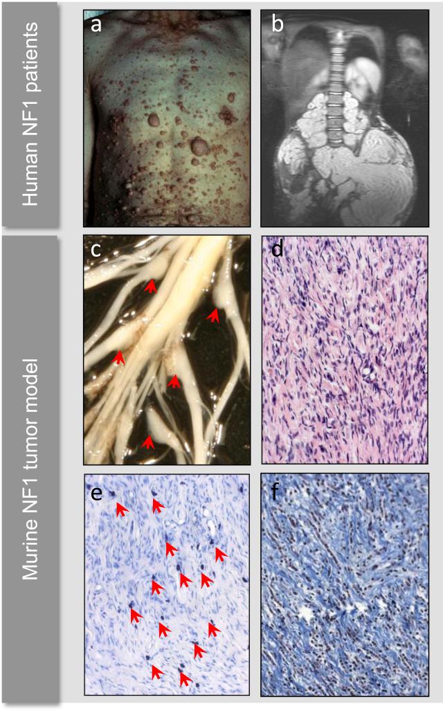
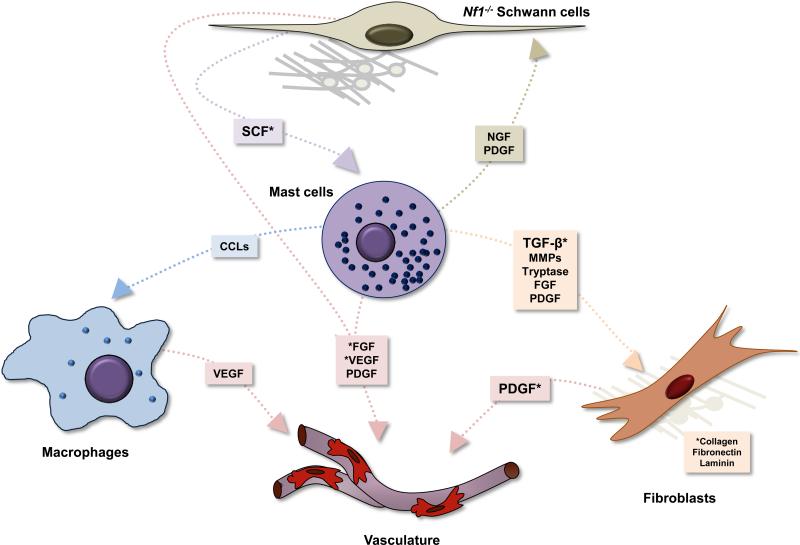
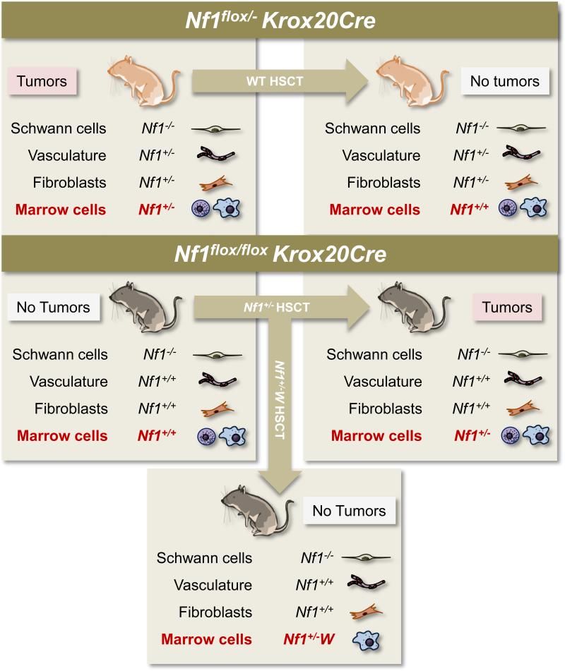
Neurofibromatosis type 1 (NF1) is a genetic disease that results from either heritable or spontaneous autosomal dominant mutations in the NF1 gene. A second-hit mutation precedes the predominant NF1 neoplasms, which include myeloid leukemia, optic glioma, and plexiform neurofibroma. Despite this requisite NF1 loss of heterozygosity in the tumor cell of origin, nontumorigenic cells contribute to both generalized and specific disease manifestations. In mouse models of plexiform neurofibroma formation, Nf1 haploinsufficient mast cells promote inflammation, accelerating tumor formation and growth. These recruited mast cells, hematopoietic effector cells long known to permeate neurofibroma tissue, mediate key mitogenic signals that contribute to vascular ingrowth, collagen deposition, and tumor growth. Thus, the plexiform neurofibroma microenvironment involves a tumor/stromal interaction with the hematopoietic system that depends, at the molecular level, on a stem cell factor/c-kit-mediated signaling axis. These observations parallel findings in other NF1 disease manifestations and are clearly relevant to medical management of these neurofibromas.


Figures

Figure 1
Figure 1. Cutaneous neurofibromas, plexiform neurofibromas, and histology
Example of cutaneous neurofibromas covering the chest and abdomen of a patient with neurofibromatosis type 1 (a). MRI of a large plexiform neurofibroma compressing the spinal column and abdominal viscera (b). Murine models of NF1-associated plexiform neurofibroma development develop enlarged dorsal root ganglia (c, red arrows), which are histologically comprised of wavy Schwann cells, numerous fibroblasts, and a granular mast cell infiltrate (d). Alcian blue deeply stains mast cell granules (e, red arrows), and trichrome stain highlights the abundant collagenization typical of neurofibromas (e). Human photograph samples reproduced with permission from the Children's Tumor Foundation: www.ctf.org.
Figure 2
Figure 2. Effect of haploinsufficiency of Nf1 on coat color and total numbers of cutaneous and peritoneal mast cells
(A) Coat color pattern of a representative mouse from each of the following genotypes: +/+;+/+, Nf1+/-;+/+, +/+;W41/W41, and Nf1+/-;W41/W41. Haploinsufficiency at Nf1partially corrects the coat color deficiency in mice homozygous for the W41 allele in a C57BL/6 genetic background. (B) Representative cytospins from peritoneal lavages stained for mast cells from individual mice of the four Nf1 and W genotypes. Peritoneal cells were stained with toluidine blue to quantify the total number of mast cells per peritoneal lavage. A higher magnification of a representative mast cell is shown in the inset of the wild-type mouse (original magnification: 3200). Bar (inset) 10 μm. Bar (far right) 30 μm. (C) Representative ear biopsies stained for cutaneous mast cells from individual mice of the four Nf1 and W genotypes. Specimens were stained with hematoxylin-eosin to assess routine histology, and with Giemsa to identify mast cells. Ear biopsies were stained with Fontana-Masson to differentiate melanin-containing cells from mast cells. Cutaneous mast cells (Giemsa-positive, Fontana-Masson–negative) were quantitated in a blinded fashion by counting the distal 5 mm of ears. Black arrows indicate Giemsa-positive mast cells, and open arrows indicate Fontana-Masson melanin–containing cells. Bar, 35 μm. © Ingram et al.,2000. Originally published in J. Exp.Med. 191: 181–188.
Figure 3
Figure 3. Hyperactive SCF/c-kit pathways in the Nf1+/- mast cell
Kit-ligand (SCF) binding at the c-kit receptor tyrosine kinase induces receptor dimerization, activates Ras to its GTP-bound conformation, and induces Ras-Raf-Mek-Erk and PI-3K-Rac-Pak-p38 signaling pathways. While Mek-Erk signals may principally mediate mast cell proliferation, PI-3K mediates survival, motility and, through its Pak-dependent crosstalk with Raf-Mek, proliferation. Nf1 accelerates the intrinsic hydrolysis of Ras-GTP to inactive Ras-GDP and serves, at least in part, to negatively regulate Mek-Erk- and PI-3K-directed pathways. Although SCF-c-kit interactions initiate other molecular events (e.g. Akt-mTOR), this schematic highlights only those thus far shown to be hyperactivate in the Nf1+/- mast cell. Dashed lines indicate multiple downstream effectors not fully detailed.
Figure 4
Figure 4. Hypothetical tumor/stromal/hematopoietic interactions in the neurofibroma microenvironment
In this tumor microenvironment model, loss of heterozygosity in Schwann cells or their precursor leads to aberrant SCF production and secretion, the ligand for the c-kit receptor tyrosine kinase. SCF induces maturation, proliferation, and recruitment of mast cells from bone marrow progenitor cells. These SCF-activated mast cells generate and release multiple inflammatory cytokines and growth factors acting on macrophages, vasculature, fibroblasts, and the tumorigenic Schwann cells. TGF-β-stimulated fibroblasts, in turn, aberrantly proliferate and produce collagen, increasing tumor bulk and pressure. Schwann cells, fibroblasts, and macrophages may all directly contribute to vascularization through various growth factors. Asterisks denote heterotypic interactions with experimental validation.
Figure 5
Figure 5. Transplant experiments in the Krox20Cre tumor model
The left column shows the genetic status of Schwann cells, vascular cells, fibroblasts, and marrow cells in two different mice genotypes: the Nf1flox/-Krox20Cre tumor model and the Nf1flox/floxKrox20Cre model, which does not develop tumors. The arrows indicate the genotype of the hematopoietic stem cells transplanted into these two different genotypes of mice. “W” represents either W41 or Wv c-kit receptor tyrosine kinase deficient mutations. The right column and bottom box show the resultant genetic status of each cell lineage, post-transplant. As indicated, only those mice with Nf1-/- Schwann cells and c-kit-competent Nf1+/- bone marrow develop tumors. HSCT: hematopoietic stem cell transplant.

References

    1. Friedman J, Gutmann DH, Maccollin M, Riccardi VM. Neurofibromatosis: phenotype, natural history, and pathogenesis. The Johns Hopkins University Press; Baltimore: 1999. p. 381.
    1. Lubs ML, Bauer MS, Formas ME, Djokic B. Lisch nodules in neurofibromatosis type 1. N Engl J Med. 1991;324:1264–6. - PubMed
    1. Riccardi VM. Neurofibromatosis: past, present, and future. N Engl J Med. 1991;324:1283–5. - PubMed
    1. Riccardi VM. Neurofibromatosis type 1 is a disorder of dysplasia: the importance of distinguishing features, consequences, and complications. Birth Defects Res A Clin Mol Teratol. 2010;88:9–14. - PubMed
    1. Akenside M. Observations on cancers. Med Trans Coll Phys Lond. 1768;1:64–92.

Publication types

MeSH terms