

Cerebellum and processing of negative facial emotions: cerebellar transcranial DC stimulation specifically enhances the emotional recognition of facial anger and sadness
- PMID: 22077643
- PMCID: PMC4234053
- DOI: 10.1080/02699931.2011.619520
Cerebellum and processing of negative facial emotions: cerebellar transcranial DC stimulation specifically enhances the emotional recognition of facial anger and sadness
Abstract
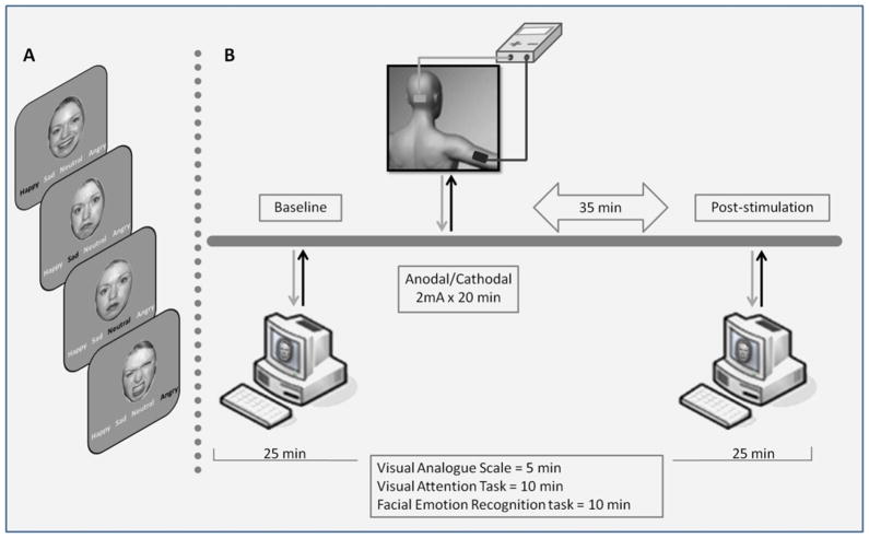
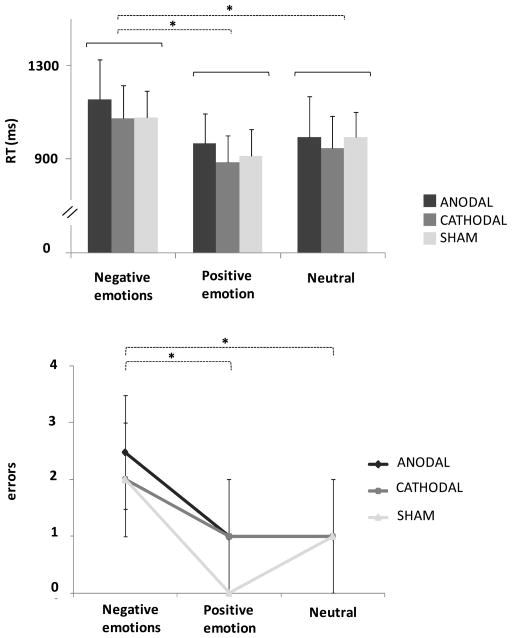
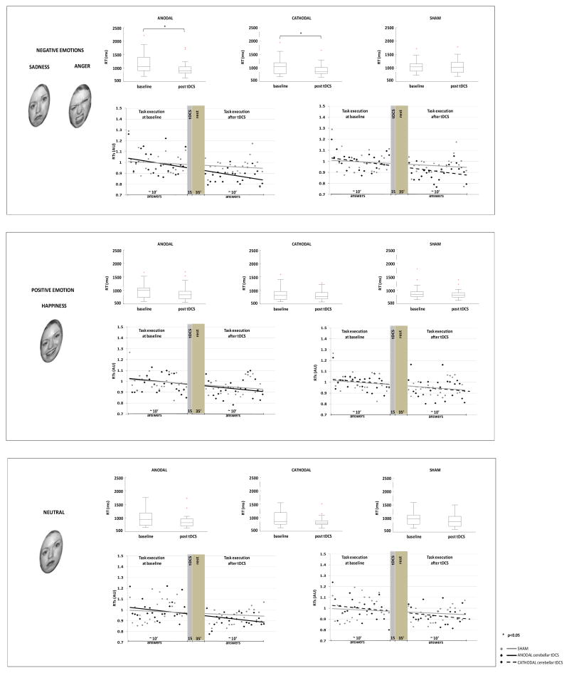
Some evidence suggests that the cerebellum participates in the complex network processing emotional facial expression. To evaluate the role of the cerebellum in recognising facial expressions we delivered transcranial direct current stimulation (tDCS) over the cerebellum and prefrontal cortex. A facial emotion recognition task was administered to 21 healthy subjects before and after cerebellar tDCS; we also tested subjects with a visual attention task and a visual analogue scale (VAS) for mood. Anodal and cathodal cerebellar tDCS both significantly enhanced sensory processing in response to negative facial expressions (anodal tDCS, p=.0021; cathodal tDCS, p=.018), but left positive emotion and neutral facial expressions unchanged (p>.05). tDCS over the right prefrontal cortex left facial expressions of both negative and positive emotion unchanged. These findings suggest that the cerebellum is specifically involved in processing facial expressions of negative emotion.
Conflict of interest statement
Roberta Ferrucci reports no financial interests or potential conflicts of interests Gaia Giannicola reports no financial interests or potential conflicts of interests Manuela Rosa reports no financial interests or potential conflicts of interests Manuela Fumagalli reports no financial interests or potential conflicts of interests Prof. Paulo S Boggio reports no financial interests or potential conflicts of interests Prof. Mark Hallett reports no financial interests or potential conflicts of interests Stefano Zago reports no financial interests or potential conflicts of interests Prof. Alberto Priori reports no financial interests or potential conflicts of interests
Figures
References
-
- Adolphs R. Neural systems for recognizing emotion. Current Opinion in Neurobiology. 2002;12(2):169–177. - PubMed
-
- Andreasen NC, O’Leary DS, Arndt S, Cizadlo T, Hurtig R, Rezai K, et al. Neural substrates of facial recognition. The Journal of Neuropsychiatry and Clinical Neurosciences. 1996;8:139–146. - PubMed
-
- Axelsson S, Kjaer I, Heiberg A, Bjornland T, Storhaug K. Neurocranial morphology and growth in Williams syndrome. European Journal of Orthodontics. 2005;27(1):32–47. - PubMed
-
- Blair RJ, Morris JS, Frith CD, Perrett DI, Dolan RJ. Dissociable neural responses to facial expressions of sadness and anger. Brain. 1999;122(5):883–893. - PubMed
MeSH terms
Grants and funding
LinkOut - more resources
Full Text Sources
Medical