

Review
doi: 10.1186/bcr2912.
Epub 2011 Nov 1.
The microenvironment in breast cancer progression: biology and implications for treatment
Affiliations
- PMID: 22078026
- PMCID: PMC3326543
- DOI: 10.1186/bcr2912
Item in Clipboard
Review
The microenvironment in breast cancer progression: biology and implications for treatment
Breast Cancer Res.
2011.
Abstract
Breast cancer comprises a heterogeneous group of malignancies derived from the ductal epithelium. The microenvironment of these cancers is now recognized as a critical participant in tumor progression and therapeutic responses. Recent data demonstrate significant gene expression and epigenetic alterations in cells composing the microenvironment during disease progression, which can be explored as biomarkers and targets for therapy. Indeed, gene expression signatures derived from tumor stroma have been linked to clinical outcomes. There is increasing interest in translating our current understanding of the tumor microenvironment to the development of novel therapies.
Figures
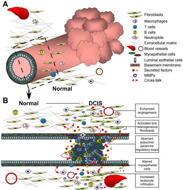
Alterations of the microenvironment from normal duct to in situ transition. (A) Schematic (transverse) view of a normal breast duct composed of a layer of luminal epithelial cells encircled by myoepithelial cells (green) and surrounded by a continuous basement membrane. Stroma containing fibroblasts, immune cells, and vasculature surrounded by the extracellular matrix maintains the normal tissue structure. (B) Longitudinal view of the normal duct and in situ carcinoma. In ductal carcinoma in situ (DCIS), epigenetically and phenotypically altered myoepithelial cells (shown as brown cells) are surrounded by a still largely continuous basement membrane. Altered myoepithelial cells in DCIS are unable to aid polarization and organize the structure of the normal duct. At the same time in the stroma, the numbers of fibroblasts and infiltrated leukocytes are increased and angiogenesis is enhanced. Cancer-associated fibroblasts (shown as yellow-green fibroblasts) and infiltrated leukocytes elevate secretion of growth factors, cytokines, chemokines, and matrix metalloproteinases (MMPs) to promote tumor progression. Potential cross-talk between cell-cell and cell-matrix interactions are aberrantly regulated by both autocrine and paracrine networks of proteolytic enzymes, cytokines, and chemokines (red arrows; not all possible interactions are indicated). Interactions between stromal and cancer cells may interact with each other via paracrine signaling rather than direct cell-cell contact.

Alterations of the microenvironment in breast cancer progression from in situ to invasive carcinoma. (A) Schematic (transverse) view of the ductal carcinoma in situ (DCIS). Although the ducts are enclosed by the altered myoepithelial cells surrounded by the basement membrane, the multiple cell types of the stroma of DCIS have dramatically changed to create a favorable tumor microenvironment. (B) Longitudinal view of the duct from DCIS to invasive ductal carcinoma transition. Invasive ductal carcinoma (IDC) is defined by degradation of the basement membrane, loss of myoepithelial cells, and invasion of epithelial cells into the stroma and vasculature. Tumor cells invade into the local environment due to the loss of the structural duct and autocrine/paracrine signaling that activated cell migration. The production of extracellular matrix-degrading proteases by the tumor cells and stromal cells is elevated during the in situ to invasive carcinoma transition, leading to destruction of the extracellular matrix such that the tumor cells can invade locally and release more secreted factors. Aberrantly secreted proteolytic enzymes, chemokines, and cytokines continue to attract leukocytes, modulate tumor remodeling, and increase tumor cell invasion to distant organs, eventually leading to metastasis.
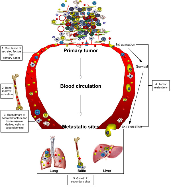
Microenvironment in the metastasis process. Metastasis is a complicated multistep process that requires cancer cells to escape from the primary tumor, survive in the circulation, seed at distant sites and grow. Each step involves stromal cell and paracrine interactions of the microenvironment. Aberrantly secreted chemokines and cytokines from the primary tumor circulate into the blood stream, creating a premetastatic niche even before tumor cell mobilization. Secreted factors functionally activate bone marrow-derived cells, which are then released into the circulation to subsequently incorporate these cells into distant organs, such as lung and liver, to create a favorable microenvironment for the cancer cell to be seeded. For the cancer cells to invade into the blood circulation, proteases are produced by bone marrow-derived cells, including macrophages and fibroblasts. Following tumor cell intravasation, a series of steps is required for the establishment of secondary tumors in the metastatic sites. Disseminated cancer cells preferentially form metastases at sites where activated bone marrow-derived cells are localized and the primary tumor has created a favorable environment at the local organ. After seeding, persistent growth of the metastatic tumor requires the establishment of a vasculature that can be possibly achieved through the production of angiogenic growth factors.
References
-
- Howlett AR, Bissell MJ. The influence of tissue microenvironment (stroma and extracellular matrix) on the development and function of mammary epithelium. Epithelial Cell Biol. 1993;2:79–89. - PubMed
-
- Petersen OW, Ronnov-Jessen L, Howlett AR, Bissell MJ. Interaction with basement membrane serves to rapidly distinguish growth and differentiation pattern of normal and malignant human breast epithelial cells. Proc Natl Acad Sci USA. 1992;89:9064–9068. doi: 10.1073/pnas.89.19.9064. A published erratum appears in Proc Natl Acad Sci USA 1993, 90:2556. - DOI - PMC - PubMed
Publication types
MeSH terms
Substances
Grants and funding
LinkOut - more resources
Full Text Sources
Other Literature Sources
Medical
