. 2012 Jan 5;422(1):144-50.
doi: 10.1016/j.virol.2011.10.008. Epub 2011 Nov 10.

Full genome analysis of a novel adenovirus from the South Polar skua (Catharacta maccormicki) in Antarctica

Affiliations

Full genome analysis of a novel adenovirus from the South Polar skua (Catharacta maccormicki) in Antarctica

Yon Mi Park et al. Virology. .

Abstract

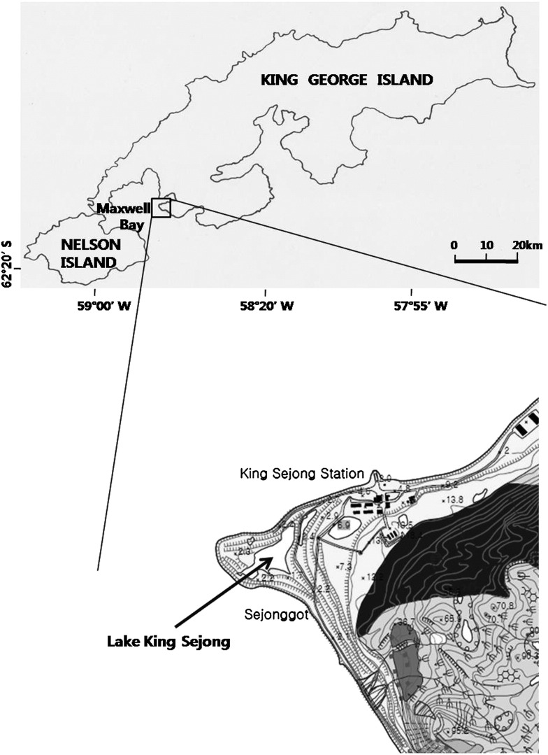
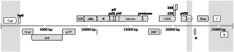
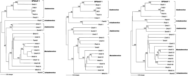
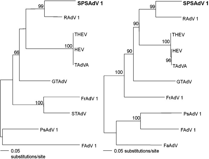
Adenoviruses have been identified in humans and a wide range of vertebrate animals, but not previously from the polar region. Here, we report the entire 26,340-bp genome of a novel adenovirus, detected by PCR, in tissues of six of nine South Polar skuas (Catharacta maccormicki), collected in Lake King Sejong, King George Island, Antarctica, from 2007 to 2009. The DNA polymerase, penton base, hexon and fiber genes of the South Polar skua adenovirus (SPSAdV) exhibited 68.3%, 75.4%, 74.9% and 48.0% nucleotide sequence similarity with their counterparts in turkey hemorrhagic enteritis virus. Phylogenetic analysis based on the entire genome revealed that SPSAdV belonged to the genus Siadenovirus, family Adenoviridae. This is the first evidence of a novel adenovirus, SPSAdV, from a large polar seabird (family Stercorariidae) in Antarctica.


Figures

Fig. 1
Fig. 1
Collection site of South Polar skua carcasses. Lake King Sejong is located southeast of King Sejong station, on King George Island, in Antarctica.
Fig. 2
Fig. 2
Genome map of South Polar skua adenovirus. The central horizontal line represents the double-stranded DNA marked at 5-kb intervals. Gray blocks show genus-common genes that can be found in every Adenoviridae genus. Sial (sialidase), hyd (hydrophobic), E3, ORF 7 and ORF 8 are genes that can be found only in Siadenovirus. Putative proteins were determined by aligning sequences of other siadenoviruses.
Fig. 3
Fig. 3
Phylogenetic trees, based on the entire amino acid sequences of the polymerase (left), penton base (middle), hexon (right) genes, generated by the neighbor-joining method. Phylogenetic relationships of SPSAdV are shown with raptor adenovirus 1 (RAdV-1, EU715130), avirulent turkey hemorrhagic enteritis virus (THEV, AY849321), hemorrhagic enteritis virus (HEV, AF074946), turkey adenovirus A (TAdV-A, AC000016), frog adenovirus 1 (FrAdV-1, AF224336), psittacine adenovirus (PsAdV, pol, EU056825; hexon, EU627198), Sulawesi tortoises adenovirus (STAdV, EU056826), great tit adenovirus (GTAdV, FJ849795), fowl adenovirus 1 (FAdV-1, U46933), falcon adenovirus (FaAdV, AY683541), duck adenovirus 1 (DAdV-1, Y09598), snake adenovirus 1 (SnAdV-1, DQ106414), ovine adenovirus 7 (OAdV-7, OAU40839), bovine adenovirus 3 (BAdV-3, AF030154), canine adenovirus 2 (CAdV-2, AC000020), porcine adenovirus A (PAdV-A, NC_005869), tree shrew adenovirus (TsAdV, NC_004453), murine adenovirus A (MAdV-A, AC000012), equine adenovirus 2 (EAdV-2, L80007), human adenovirus 1 (HAdV-1, AC000017), human adenovirus 3 (HAdV-3, DQ086466), human adenovirus 4 (HAdV-4, AY458656), human adenovirus 8 (HAdV-8, AB448769), human adenovirus 12 (HAdV-12, X73487), human adenovirus 40 (HAdV-40, NC_001454), simian adenovirus 21 (SAdV-21, AC000010) and white sturgeon adenovirus (WsAdV, AY082701). Branch lengths are proportional to the number of amino acid substitutions, while vertical distances are for clarity only. The numbers at each node are bootstrap probabilities (expressed as percentages), as determined for 1000 iterations by PAUP version 4.0b.
Fig. 4
Fig. 4
Phylogenetic trees, based on the partial nucleotide sequences of the polymerase (275 bp, left) and hexon (608 bp, right) genes, generated by the neighbor-joining method. Phylogenetic relationships of SPSAdV are shown with other Siadenovirus and Aviadenovirus, including raptor adenovirus 1 (RAdV-1, EU715130), avirulent turkey hemorrhagic enteritis virus (THEV, AY849321), hemorrhagic enteritis virus (HEV, AF074946), turkey adenovirus A (TAdV-A, AC000016), frog adenovirus 1 (FrAdV-1, AF224336), psittacine adenovirus (PsAdV, pol gene: EU056825; hexon gene: EU627198), Sulawesi tortoises adenovirus (STAdV, EU056826), great tit adenovirus (GTAdV, FJ849795), fowl adenovirus 1 (FAdV-1, U46933), falcon adenovirus (FaAdV, AY683541). Branch lengths are proportional to the number of nucleotide substitutions, while vertical distances are for clarity only. The numbers at each node are bootstrap probabilities (expressed as percentages), as determined for 1000 iterations by PAUP version 4.0b.

References

    1. Aggarwal N., Mittal S.K. Sequence analysis of porcine adenovirus type 3 E1 region, pIX and pIVa2 genes, and two novel open reading frames. Intervirology. 2000;43(1):6–12. - PubMed
    1. Altschul S.F., Gish W., Miller W., Myers E.W., Lipman D.J. Basic local alignment search tool. J. Mol. Biol. 1990;215(3):403–410. - PubMed
    1. Austin F.J., Webster R.G. Evidence of ortho- and paramyxoviruses in fauna from Antarctica. J. Wildl. Dis. 1993;29(4):568–571. - PubMed
    1. Beach N.M., Duncan R.B., Larsen C.T., Meng X.J., Sriranganathan N., Pierson F.W. Comparison of 12 turkey hemorrhagic enteritis virus isolates allows prediction of genetic factors affecting virulence. J. Gen. Virol. 2009;90(Pt 8):1978–1985. - PubMed
    1. Benkö M., Harrach B., Russell W.C. Family Adenoviridae. In: Van Regenmortel M.H.V., Fauquet C.M., Bishop D.H.L., Carstens E., Estes M., Lemon S., Maniloff J., Mayo M.A., McGeoch D., Pringle C., Wickner R., editors. Virus Taxonomy. VIIth Report of the International Committee on Taxonomy of Viruses Academic Press; New York: 2000.

Publication types

Substances