Review
. 2011 Nov 11;147(4):742-58.
doi: 10.1016/j.cell.2011.10.033.

Programmed cell death in animal development and disease

Affiliations
Review

Programmed cell death in animal development and disease

Yaron Fuchs et al. Cell. .

Erratum in

  • Cell. 2011 Dec 23;147(7):1640

Abstract

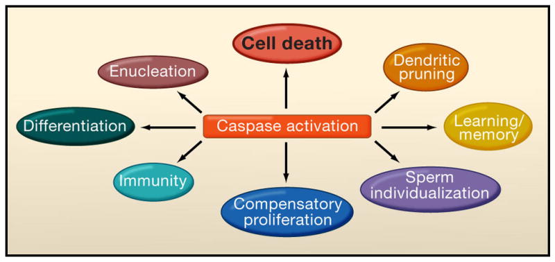
Programmed cell death (PCD) plays a fundamental role in animal development and tissue homeostasis. Abnormal regulation of this process is associated with a wide variety of human diseases, including immunological and developmental disorders, neurodegeneration, and cancer. Here, we provide a brief historical overview of the field and reflect on the regulation, roles, and modes of PCD during animal development. We also discuss the function and regulation of apoptotic proteins, including caspases, the key executioners of apoptosis, and review the nonlethal functions of these proteins in diverse developmental processes, such as cell differentiation and tissue remodeling. Finally, we explore a growing body of work about the connections between apoptosis, stem cells, and cancer, focusing on how apoptotic cells release a variety of signals to communicate with their cellular environment, including factors that promote cell division, tissue regeneration, and wound healing.


Figures

Figure 1
Figure 1. Evolutionary conservation of the core apoptotic machinery
A comparison between the apoptotic pathways in C. elegans, Drosophila and mammals reveals conservation and expansion of the apoptotic pathway during evolution. A. In C. elegans apoptotic signals regulate the interplay between Egl-1 (BH3-only homology) and CED-9 (Bcl-2 family homologue), liberating CED-4 (Apaf-1 homologue) to activate CED-3 (caspase-9 homologue) for programmed death of 131 cells. B. In Drosophila, many different signaling pathways regulate both the IAP antagonists Reaper, Hid, and Grim (RHG) and the apoptosome proteins Ark (Apaf-1 homologue) and Dronc (caspase-9 homologue). On the one hand, this causes the ubiquitin-mediated degradation of DIAP1 and de-repression of caspases, and on the other hand it enables Dronc (caspase-9 homologue) to associate with Ark, creating active apoptosomes and activation of the effector caspases DrICE and Dcp1. Both pathways are required for efficient caspase activation and are coordinately regulated, in analogy with driving a car with “gas” and “brake”. However, removal of the “brakes” is necessary for the efficient induction of apoptosis in vivo and often initiates it. The P35 protein can specifically inhibit the activity of Dcp-1 and DrICE. C. In mammals, the balance between pro-apoptotic and anti-apoptotic Bcl-2 family members is a key factor in the commitment to apoptosis by regulating the release of cytochrome c and IAP antagonists from mitochondria. Binding of cytochrome c to Apaf-1 promotes apoptosome assembly, which recruits and activates caspase-9. IAP antagonists liberate caspases from the inhibition of IAPs, most notably XIAP (X-linked inhibitor of apoptosis), which targets both initiator and effector caspases. The XIAP-antagonist ARTS is localized to the mitochondrial outer membrane and acts prior to the release of cytochrome c, Smac and other proteins released from the mitochondrial inter-membrane space. Homologues proteins (by either function or sequence) are similarly illustrated.
Figure 2
Figure 2. Functions of PCD during development
(A) PCD regulates proper structure sculpting by eliminating interdigital-webbings. (B) During Drosophila metamorphosis nearly all larval structures are destroyed such as the Salivary glands (SG), Muscles (M), midgut (MG) and hindgut (HG). (depicted by purple), whereas novel structures are raised from undifferentiated cells termed imaginal discs (depicted by various colors). The locations and developmental fates of the imaginal discs are similarly illustrated. PCD also controls cell number for example by deleting cells which fail to partner (C) and eliminates dangerous and abnormal cells such as autoreactive lymphocytes (D).
Figure 3
Figure 3. Integration of different signaling pathways by RHG proteins
The genes encoding Reaper, Hid and Grim (RHG) are the targets for regulation of many signaling pathways that influence the decision between cell death and survival. The transcriptional control region of RHG genes, exemplified here by reaper, contains binding sites for many different transcription factors that are the downstream targets of different signaling pathways, including for the steroid hormone ecdysone, patterning signals and Hox genes, and DNA-damage/p53. Examples for both activators (green) and repressors (red) have been described. The steroid hormone ecdysone can either induce or repress reaper transcription depending at different stages of development. RHG genes are clustered and, to some extent, co-regulated at the transcriptional level, and the locus can be silenced by histone-modifying enzymes and Polycomb.
Figure 4
Figure 4. Apoptosis induced compensatory proliferation (CP) in various organisms
In different model organisms apoptosis and caspase activity have been observed to induce secretion of mitogenic factors, thereby promoting hyperplastic overgrowth or tissue regeneration. Mitogenic factors are indicated in pink and question marks indicate uncertainty. A. In Drosophila inhibition of caspases by P35 renders cells in an “undead” state unable to complete apoptosis. This results in the activation of p53 and JNK, triggering the release of the Wg and Dpp mitogens, thereby promoting hyperplasic overgrowth. B. In Drosophila, temporal and spatial apoptosis (in-dependently of p35) induces tissue regeneration via compensatory proliferation by secretion of Wg. A dP53/JNK positive feedback loop is essential for the apoptotic response. C. In Drosophila differentiating neurons induce a different compensatory proliferation pathway, via hedghog (Hh), in a manner requiring both DrICE and Dcp-1. Hh stimulates the proliferation of non-neuronal cells. D. In Hydra, head regeneration post midgut bisection is dependent upon caspase activity, where apoptotic cells secrete Wnt3 promoting CP. E. In newts and planaria, amputation is characterized by apoptosis and caspase activity in the wound site. However it still unknown whether this apoptotic response is responsible for the release of Wnt and Hh. F. In Xenopus, amputation of the tail results in caspase activity, whereas inhibition of caspase-9 and 3 prohibits cell proliferation and the regenerative process. It remains to established whether this form of CP is mediated by Wnt signaling. G. In mice, wound repair and liver regeneration is dependant upon caspase -3 and -7 which are necessary for proper induction of these processes. Caspase-3 mediates the proteolytic processing of iPLA2, which in turn produces archidonic acid the precursor of PGE2, a known stimulator of stem cell proliferation, tissue regeneration and wound repair.
Figure 5
Figure 5. Non-apoptotic function of caspases
In addition to their central role in apoptosis, caspases are involved in many other vital process including: differentiation, enucleation, pruning of axons and dendrites, sperm differentiation, immunity, compensatory proliferation and even learning and memory.

Similar articles

Cited by

References

    1. Aballay A, Ausubel FM. Programmed cell death mediated by ced-3 and ced-4 protects Caenorhabditis elegans from Salmonella typhimurium-mediated killing. Proc Natl Acad Sci U S A. 2001;98:2735–2739. - PMC - PubMed
    1. Abdelwahid E, Pelliniemi LJ, Jokinen E. Cell death and differentiation in the development of the endocardial cushion of the embryonic heart. Microsc Res Tech. 2002;58:395–403. - PubMed
    1. Abdelwahid E, Yokokura T, Krieser RJ, Balasundaram S, Fowle WH, White K. Mitochondrial disruption in Drosophila apoptosis. Dev Cell. 2007;12:793–806. - PubMed
    1. Abraham MC, Lu Y, Shaham S. A morphologically conserved nonapoptotic program promotes linker cell death in Caenorhabditis elegans. Dev Cell. 2007;12:73–86. - PubMed
    1. Abraham MC, Shaham S. Death without caspases, caspases without death. Trends Cell Biol. 2004;14:184–193. - PubMed

Publication types

LinkOut - more resources