. 2011 Nov 11;147(4):868-80.
doi: 10.1016/j.cell.2011.09.051.

CD14 controls the LPS-induced endocytosis of Toll-like receptor 4

Affiliations

CD14 controls the LPS-induced endocytosis of Toll-like receptor 4

Ivan Zanoni et al. Cell. .

Abstract

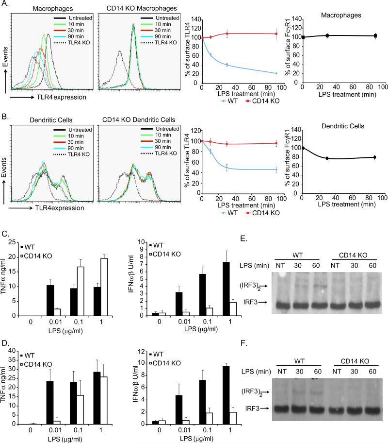
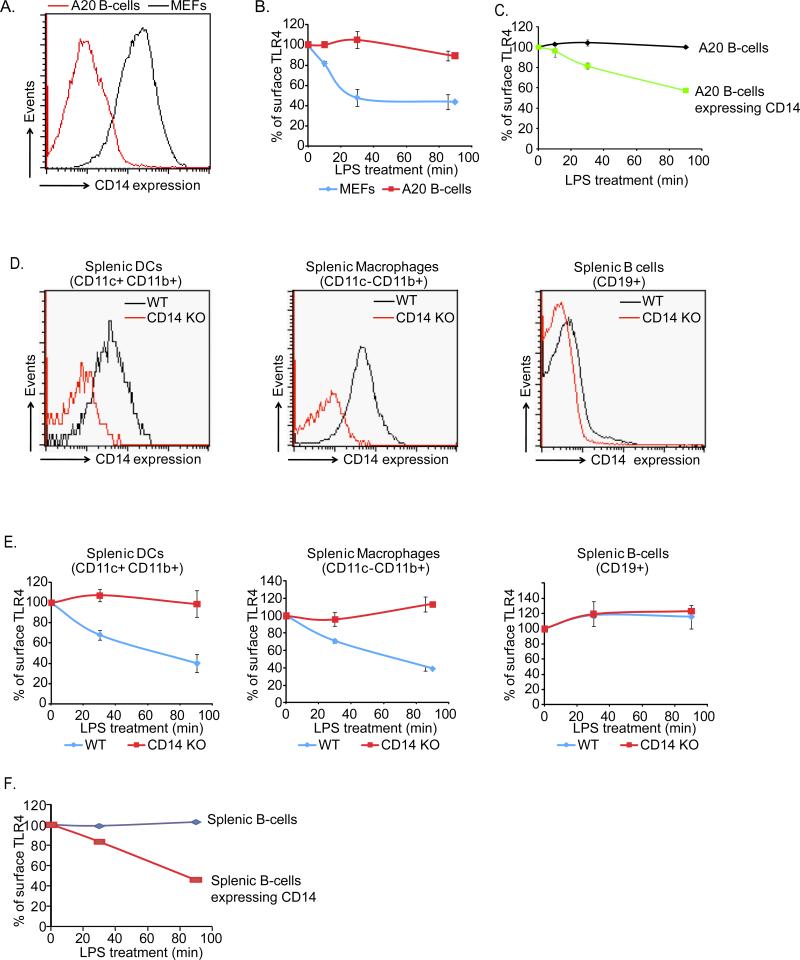
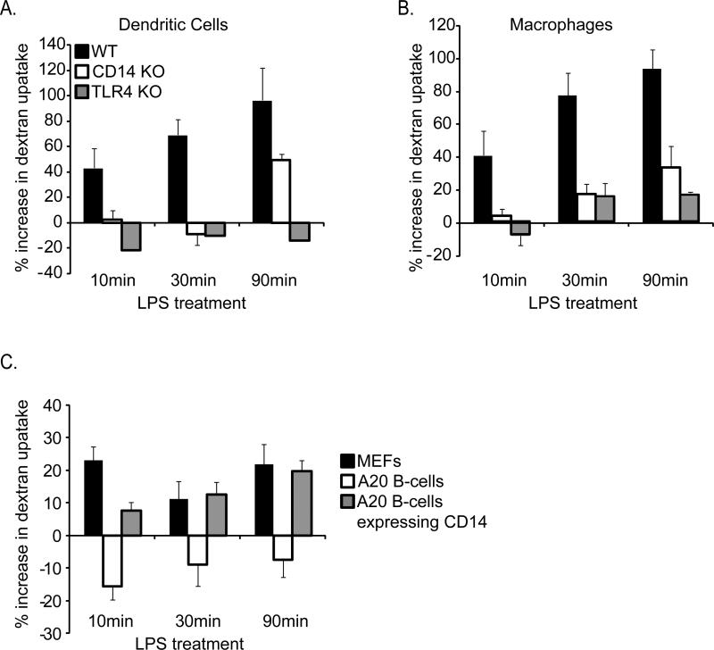
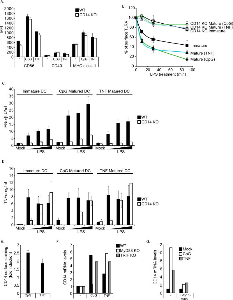
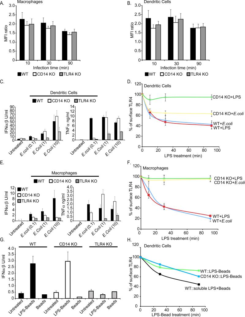
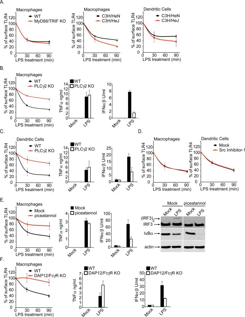
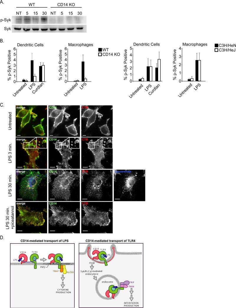
The transport of Toll-like Receptors (TLRs) to various organelles has emerged as an essential means by which innate immunity is regulated. While most of our knowledge is restricted to regulators that promote the transport of newly synthesized receptors, the regulators that control TLR transport after microbial detection remain unknown. Here, we report that the plasma membrane localized Pattern Recognition Receptor (PRR) CD14 is required for the microbe-induced endocytosis of TLR4. In dendritic cells, this CD14-dependent endocytosis pathway is upregulated upon exposure to inflammatory mediators. We identify the tyrosine kinase Syk and its downstream effector PLCγ2 as important regulators of TLR4 endocytosis and signaling. These data establish that upon microbial detection, an upstream PRR (CD14) controls the trafficking and signaling functions of a downstream PRR (TLR4). This innate immune trafficking cascade illustrates how pathogen detection systems operate to induce both membrane transport and signal transduction.


Figures

Figure 1
Figure 1. CD14 is required for LPS-induced TLR4 endocytosis
WT or CD14-deficient (KO) mouse BMDM(A) or DCs (B) were untreated or treated with LPS (1μg/ml) for the times indicated. Flow cytometry was then used to examine receptor endocytosis by determining the surface levels of the endogenous proteins indicated. The third and forth panels in A and B represent the mean fluorescence intensity (MFI) of specific receptor staining at each time point. C, D; BMDM (C) or DCs (D) were treated with the concentrations of LPS indicated for 18 hours and the amounts of secreted cytokines were determined. E, F; BMDM (E) or DCs (F) were treated with LPS (1μg/ml) for the times indicated and the presence of active (dimerized) IRF3 in cell extracts was determined by native PAGE. See also Figure S1.
Figure 2
Figure 2. The natural expression profile of CD14 determines which cells undergo LPS-induced TLR4 endocytosis
A, The cells indicated were examined for the expression of surface levels of endogenous CD14 by flow cytometry. B, MEFs and A20 B-cells were treated with LPS (1μg/ml) for the times indicated and TLR4 endocytosis was monitored by flow cytometry. Shown are the MFI of specific TLR4 surface staining at each time point indicated. C, A20 B-cells were transiently transfected with a plasmid encoding CD14 and treated with LPS (1μg/ml) for the times indicated before TLR4 endocytosis was examined by flow cytometry. D, DCs, BMDM or B-cells were isolated from the spleen of mice and examined for the expression of surface levels of CD14 by flow cytometry. E, Mice were injected with LPS (50μg) and at the times indicated spleens were isolated and BMDM, DCs and B-cells were examined for surface levels of TLR4 by flow cytometry. F, Splenic B-cells were transfected with a plasmid encoding CD14 and treated for the indicated times with LPS (1μg/ml) before TLR4 endocytosis was examined by flow cytometry. See also Figure S2.
Figure 3
Figure 3. CD14 is required for LPS-induced macropinocytosis
Macropinocytosis was measured in DCs (A) or BMDM (B) by flow cytometry. Cells were untreated or treated for the times indicated with fluorescent dextrans (1mg/ml) in the presence or absence of LPS (1μg/ml). Cell associated fluorescence intensity was then compared between LPS-treated and untreated cell populations to determine changes from the steady state levels of macropinocytosis. Note that both CD14 and TLR4 are required for LPS-induced macropinocytosis. C, MEFs or A20 B-cells were untreated or treated for the times indicated with fluorescent dextrans (1mg/ml) in the presence or absence of LPS (1μg/ml). Cell associated fluorescence intensity was then compared between LPS-treated and untreated populations of cells to determine changes from the steady state levels of macropinocytosis.
Figure 4
Figure 4. The CD14-dependent endocytosis pathway is upregulated during DC maturation
A, DCs were untreated or treated for 18 hours with CpG DNA (1μM) or TNFα (100pg/ml). DC maturation was then assessed by flow cytometry by determining the increase in surface staining of CD86, CD40 or MHC class II. B, Immature DCs or DCs matured for 18 hours with CpG DNA (1μM) or TNFα (100pg/ml) were treated with LPS (1μg/ml) and TLR4 endocytosis was measured by flow cytometry at the times indicated. Displayed are the MFIs of specific surface TLR4 staining at each time point. C, D; Immature DCs or DCs matured for 18 hours with CpG DNA (1μM) or TNFα (100pg/ml) were treated with LPS at the concentrations indicated and the production of the indicated cytokines were measured after 18 hours. E, DCs were either untreated or treated for 18 hours with CpG DNA (1μM) or TNFα (100pg/ml) and the affect of these stimuli on the cell surface levels of CD14 was assessed by flow cytometry. F, DCs of the genotypes indicated were processed as described in (E). After 18 hours, CD14 mRNA levels were determined by qPCR. G, DCs were stimulated with the ligands indicated as described in (E) in the presence of absence of the NF-κB inhibitor Bay11-7085 and then processed to measure CD14 expression by qPCR.
Figure 5
Figure 5. The requirement of CD14 for TLR4 endocytosis can be bypassed with phagocytic cargo
A, B; The cells indicated were treated for the times indicated with fluorescent bacteria at a multiplicity of infection (MOI) of 10. Phagocytosis was then measured at each time point by determining the mean fluorescence intensity (MFI) of the cell populations by flow cytometry. C, DCs were treated with E.coli at the MOIs indicated on the x-axis, and the production of the indicated cytokines were measured after 18 hours. D, DCs were treated with E.coli at an MOI of 10 or LPS (1μg/ml) and TLR4 endocytosis was measured by flow cytometry at the times indicated. Displayed are the MFIs of specific surface TLR4 staining at each time point. E, BMDM were treated with E.coli at the MOIs indicated on the x-axis, and the production of the cytokines indicated were measured. F, BMDM were treated with E.coli at an MOI of 10 or LPS (1μg/ml) and TLR4 endocytosis was measured by flow cytometry at the times indicated. Displayed are the MFIs of specific surface TLR4 staining at each time point. G, DCs were treated with either LPS-coated latex beads or uncoated beads and the production of the cytokines indicated were measured. H, DCs were treated with either LPS-coated latex beads or uncoated beads in the presence of soluble LPS and TLR4 endocytosis was measured by flow cytometry at the times indicated. Displayed are the MFIs of specific surface TLR4 staining at each time point. See also Figure S3.
Figure 6
Figure 6. Role of Syk and PLCγ2 in regulating the CD14-dependent endocytosis of TLR4
A, Immortal BMDM or primary and DCs of the genotypes indicated were either untreated or treated with LPS (1μg/ml) for the times indicated. Flow cytometry was then used to examine receptor endocytosis by determining the surface levels of the endogenous TLR4. Shown are the MFIs of specific TLR4 surface staining at each time point indicated. B, C, BMDM (B) or DCs (C) of the genotypes indicated were treated with 10ng/ml LPS for the times indicated and the rate of TLR4 endocytosis was assessed by flow cytometry (first panel). Second and third panels, cells were stimulated with 10ng/ml LPS for 18 hours and the amounts of secreted cytokines were determined. D, The cells indicated were treated with the Src inhibitor in the presence or absence of LPS (10ng/ml) and the rate of TLR4 endocytosis was monitored by flow cytometry. E, BMDM were treated with the Syk inhibitor piceatannol (75μM) and were then stimulated with 10ng/ml LPS. At the times indicated, the rate of TLR4 endocytosis was assessed by flow cytometry (first panel). Second and third panels, cells were stimulated in the presence or absence of piceatannol with 10ng/ml LPS for 18 hours and the amounts of secreted cytokines were determined. Fourth panel, BMDM were treated with LPS (1μg/ml) for 30 min in the presence or absence of piceatannol (75μM). The presence of dimerized IRF3 and IκBα was assessed by western analysis. F, BMDM of the genotypes indicated were treated with 10ng/ml LPS for either 30 or 90 minutes. The rate of TLR4 endocytosis was assessed by flow cytometry (first panel). Second and third panels, cells were stimulated with 10ng/ml LPS for 18 hours and the amounts of secreted cytokines were determined. See also Figures S4.
Figure 7
Figure 7. Syk is activated by LPS by a process dependent on CD14 but independent of TLR4
A, BMDM of the genotypes indicated were stimulated with LPS (100ng/ml). At the times indicated, the presence of phospho-Syk was examined by western blot. B, BMDM and DCs of the genotypes indicated were stimulated with either LPS or curdlan and the presence of phospho-Syk was detected by flow cytometry. C, BMDM were stimulated with LPS (100ng/ml) for the times indicated and processed for confocal microscopy to detect the presence of CD14, Cholera Toxin B (CTB) or dextran loaded lysosomes. All images for all panels are representative of at least three independent experiments where over 500 cells were examined per condition and >95% of the cells displayed similar staining. D, Model depicting a cascade of transport events that is mediated by CD14 in order to promote TLR4 signaling. CD14 first captures and transports LPS to the plasma membrane localized complex of TLR4 and MD2, which signals through the TIRAP-MyD88 adaptors to activate inflammatory cytokine expression. CD14 then transports TLR4 to endosomes by a process mediated by Syk and PLCγ2, where TRAM-TRIF signaling can lead to the expression of IFNs. See also Figure S5.

References

    1. Akira S, Takeda K. Toll-like receptor signalling. Nat Rev Immunol. 2004;4:499–511. - PubMed
    1. Barbalat R, Lau L, Locksley RM, Barton GM. Toll-like receptor 2 on inflammatory monocytes induces type I interferon in response to viral but not bacterial ligands. Nat Immunol. 2009;10:1200–1207. - PMC - PubMed
    1. Barton GM, Kagan JC. A cell biological view of Toll-like receptor function: regulation through compartmentalization. Nat Rev Immunol. 2009;9:535–542. - PMC - PubMed
    1. Botelho RJ, Teruel M, Dierckman R, Anderson R, Wells A, York JD, Meyer T, Grinstein S. Localized biphasic changes in phosphatidylinositol-4,5-bisphosphate at sites of phagocytosis. J Cell Biol. 2000;151:1353–1368. - PMC - PubMed
    1. Cheong C, Matos I, Choi JH, Dandamudi DB, Shrestha E, Longhi MP, Jeffrey KL, Anthony RM, Kluger C, Nchinda G, et al. Microbial stimulation fully differentiates monocytes to DC-SIGN/CD209(+) dendritic cells for immune T cell areas. Cell. 143:416–429. - PMC - PubMed

Publication types

MeSH terms