

Toxicity and cosmesis outcomes after single fraction partial breast irradiation in early stage breast cancer
- PMID: 22079051
- PMCID: PMC3229442
- DOI: 10.1186/1748-717X-6-155
Toxicity and cosmesis outcomes after single fraction partial breast irradiation in early stage breast cancer
Abstract
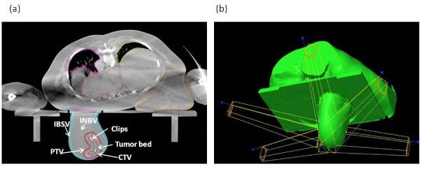
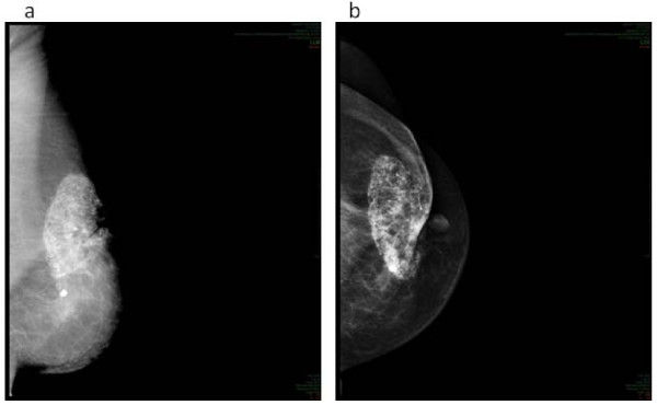
Background: To report the clinical outcome after a Single Shot 3D-CRT PBI (SSPBI) in breast cancer patients after conservative surgery (ClinicalTrials.gov Identifier: NCT01316328).
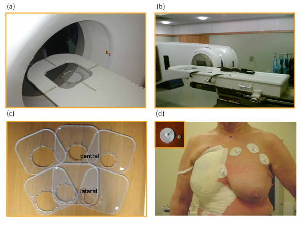
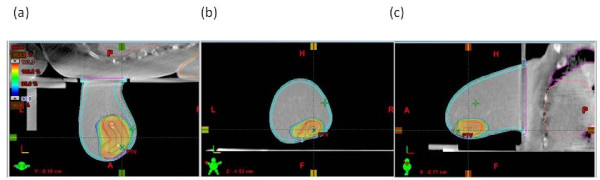
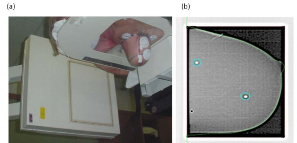
Methods: A dose of 18 Gy (in the first 4 patients) and 21 Gy (in the remaining 60 patients) was prescribed in a single session and delivered to the index area (i.e. the area of breast including the primary tumor bed and the surrounding tissue) using 3D-CRT with patients in prone position. Acute and late toxicity was assessed using the National Cancer Institute's CTC for Adverse Events. Cosmesis was defined based on modified Harvard criteria. Differences between dosimetric or clinical parameters of patients with/without G2 or more late toxicity or unsatisfactory (poor or fair) cosmetic outcome were evaluated with the Mann-Whitney test. Odds ratios and 95% confidence interval were calculated for cosmesis and fibrosis. Univariate and multivariate analyses(UVA/MVA) were used to determine covariates associated with an increase in fibrosis or fat necrosis rate.
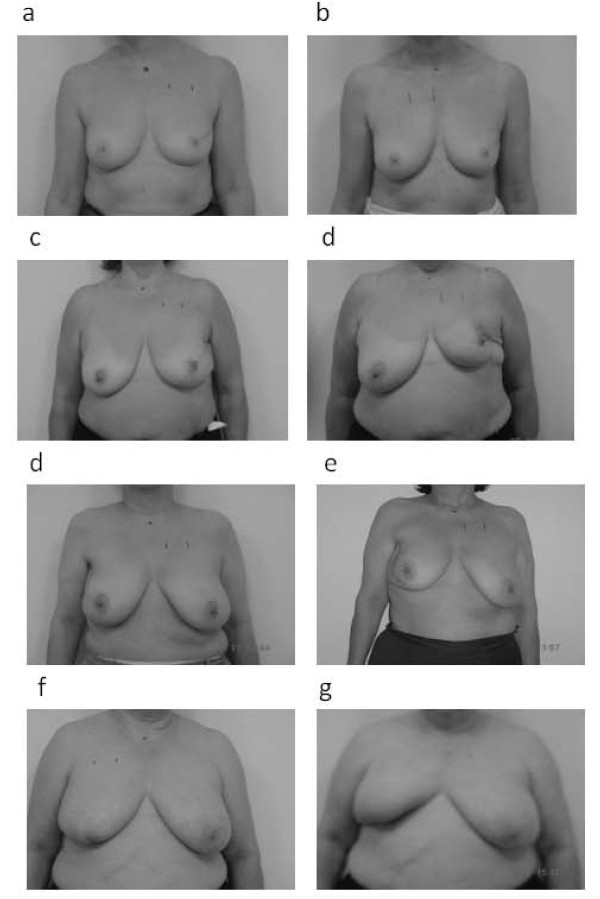
Results: Sixty four patients were enrolled. With a median follow-up of 3 years, G2 and G3 subcutaneous fibrosis was detected in 20(31%) and in 8(13%) patients, and ≥G2 fat necrosis was observed in 2(3%) patients. Good to excellent, fair and poor cosmesis was observed in 38(59%), 23(36%) and 3(5%) patients, respectively. Based on UVA, the breast volume receiving more than 21 Gy (V21 Gy) was found to be a predictor of the ≥G1 or ≥G2 fibrosis/fat necrosis. Based on MVA, V21 Gy was confirmed as a predictor for ≥G1 fibrosis/fat necrosis, the results correlated as a trend for ≥G2. Cosmesis was correlated with whole breast (WB) mean dose (p=0.030).
Conclusion: Our choice of a single dose of 21 Gy significantly increased the treatment related toxicity. However, this should not discourage novel SSPBI approaches with lower equivalent doses.
Figures
References
-
- Fisher ER, Anderson S, Redmond C, Fisher B. Ipsilateral breast tumor recurrence and survival following lumpectomy and irradiation: pathological findings from NASBP protocol B-06. Semin Surg Oncol. 1992;8:161–166. - PubMed
-
- Schnitt SJ, Abner A, Gelman R, Connolly JL, Recht A, Duda RB, Eberlein TJ, Mayzel K, Silver B, Harris JR. The relationship between microscopic margins of resection and the risk of local recurrence in patients with breast cancer treated with breast-conserving surgery and radiation therapy. Cancer. 1994;74:1746–1751. doi: 10.1002/1097-0142(19940915)74:6<1746::AID-CNCR2820740617>3.0.CO;2-Y. - DOI - PubMed
-
- R. The relationship between microscopic margins of resection and the risk of local recurrence in patients with breast cancer treated with breast-conserving surgery and radiation therapy. Cancer. 1994;74:1746–1751. doi: 10.1002/1097-0142(19940915)74:6<1746::AID-CNCR2820740617>3.0.CO;2-Y. conservatively treated.Eur.J.Cancer 2001, 37:2178-2183. - DOI - PubMed
Publication types
MeSH terms
Associated data
LinkOut - more resources
Full Text Sources
Medical
Research Materials
