

Subject-specific changes in brain white matter on diffusion tensor imaging after sports-related concussion
- PMID: 22079073
- PMCID: PMC3254806
- DOI: 10.1016/j.mri.2011.10.001
Subject-specific changes in brain white matter on diffusion tensor imaging after sports-related concussion
Abstract
Background and purpose: Current approaches to diffusion tensor imaging (DTI) analysis do not permit identification of individual-level changes in DTI indices. We investigated the ability of wild bootstrapping analysis to detect subject-specific changes in brain white matter (WM) before and after sports-related concussion.
Materials and methods: A prospective cohort study was performed in nine high school athletes engaged in hockey or football and six controls. Subjects underwent DTI pre- and postseason within a 3-month interval. One athlete was diagnosed with concussion (scanned within 72 h), and eight suffered between 26 and 399 subconcussive head blows. Fractional anisotropy (FA) and mean diffusivity (MD) were measured in each WM voxel. Bootstrap samples were generated, and a permuted t test was used to compare voxel-wise FA/MD changes in each subject pre- vs. postseason.
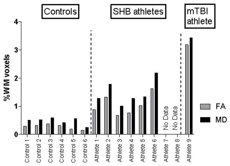
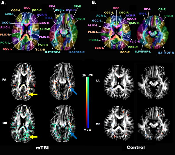
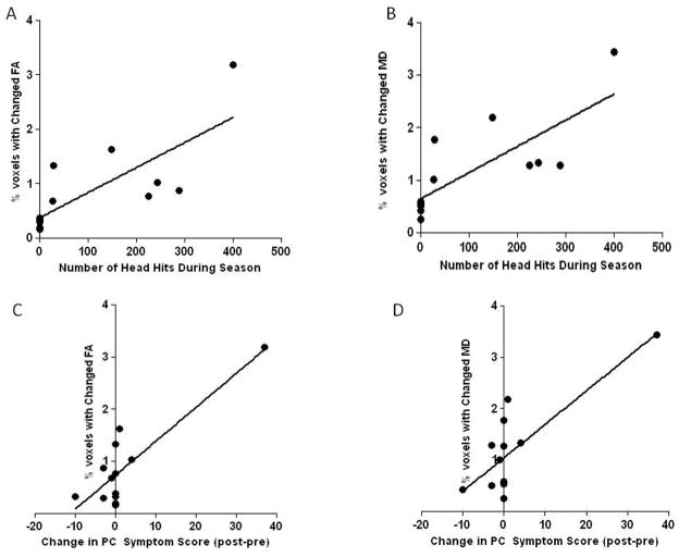
Results: The percentage of WM voxels with significant (p<.05) pre-post FA changes was highest for the concussion subject (3.2%), intermediary for those with subconcussive head blows (mean 1.05%±.15%) and lowest for controls (mean 0.28%±.01%). Similarly, the percentage of WM voxels with significant MD changes was highest for the concussion subject (3.44%), intermediary for those with subconcussive head blows (mean 1.48%±.17%) and lowest for controls (mean 0.48%±.05%). Significantly changed FA and MD voxels colocalized in the concussion subject to the right corona radiata and right inferior longitudinal fasciculus.
Conclusions: Wild bootstrap analysis detected significantly changed WM in a single concussed athlete. Athletes with multiple subconcussive head blows had significant changes in a percentage of their WM that was over three times higher than controls. Efforts to understand the significance of these WM changes and their relationship to head impact forces appear warranted.
Copyright © 2012 Elsevier Inc. All rights reserved.
Figures

References
-
- Faul M, Wald MM, Coronado VG. Traumatic Brain Injury in the United States: Emergency Department Visits, Hospitalizations and Deaths 2002–2006. Centers for Disease Control and Prevention, National Center for Injury Prevention and Control; Atlanta, GA: 2010.
-
- Thurman DJ, Branche CM, Sniezek JE. The epidemiology of sports-related traumatic brain injuries in the United States: recent developments. J Head Trauma Rehab. 1998;13(2):1–8. - PubMed
-
- Bazarian JJ, Cernak I, Noble-Haeusslein L, Potolicchio S, Temkin N. Long-term neurologic outcomes after traumatic brain injury. J Head Trauma Rehab. 2009;24(6):439–51. - PubMed
-
- Omalu BI, DeKosky ST, Minster RL, Kamboh MI, Hamilton RL, Wecht CH. Chronic traumatic encephalopathy in a National Football League player. Neurosurgery. 2005;57(1):128–34. - PubMed
-
- Povlishock JT, Katz DI. Update of neuropathology and neurological recovery after traumatic brain injury. J Head Trauma Rehab. 2005;20(1):76–94. - PubMed
Publication types
MeSH terms
Grants and funding
LinkOut - more resources
Full Text Sources
Other Literature Sources
Medical
