Review
. 2011 Dec;13(12):992-6.
doi: 10.1016/j.jfms.2011.08.003. Epub 2011 Nov 10.

Cervical dermoid sinus in a cat: case presentation and review of the literature

Affiliations
Review

Cervical dermoid sinus in a cat: case presentation and review of the literature

Jamie M Fleming et al. J Feline Med Surg. 2011 Dec.

Abstract

A 6-month-old female spayed domestic shorthair cat was presented for evaluation of a focal subcutaneous swelling on the dorsal neck at the level of atlas. The magnetic resonance imaging and surgical treatment of a dermoid sinus associated with the cervical vertebrae is described. To the authors' knowledge, a dermoid sinus in this location has not been described previously in the cat. The prognosis following surgical resection appears favorable.


Figures

Fig 1.
Fig 1.
Lateral radiograph of the cervical vertebrae (a). The intervertebral disc spaces of C2—C6 are narrowed which gives the appearance that the vertebral bodies C2 through C6 are fused. Ventrodorsal radiograph of the cervical vertebrae. There is scoliosis which is most severe at C2—C3 and C3—C4 (b).
Fig 2.
Fig 2.
A sagittal T1W FLAIR image of the brain and cranial spinal cord. The palpable subcutaneous lesion is observed in the subcutaneous fat. There is a visible depression in the epaxial muscles as the lesion penetrates the muscle. The lesion extends deep to the muscle to level of the dorsal arch of the atlas (white arrow).
Fig 3.
Fig 3.
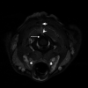
A 3D FSPGR axial image at the level of the cranial aspect of the atlas. The 3D FSPGR was performed utilizing a chemical fat saturation pulse. There is a midline defect in the dorsal arch of the atlas (white arrowhead). In comparison to the muscle, the palpable subcutaneous lesion is visible as a hyperintense structure (short white arrow). There is an expansion of the dorsal subarachnoid space (long white arrow).
Fig 4.
Fig 4.
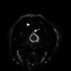
T2W axial image at the level of the atlas. The palpable subcutaneous lesion is observed in the epaxial muscles overlying the atlas (white arrowhead). There is a deviation of dorsal meninges toward the defect in the arch of the atlas which results in a triangular shape of the subarachnoid space in the axial plane (white arrow).

References

    1. Icenogle DA, Kaplan AM. A review of congenital neurologic malformations. Clin Pediatr (Phila) 1981; 20: 565–76. - PubMed
    1. Kasa F, Kasa G, Kussinger S. [Dermoid sinus in a Rhodesian Ridgeback. Case report]. Tierarztl Prax 1992; 20: 628–31. - PubMed
    1. Pratt JN, Knottenbelt CM, Welsh EM. Dermoid sinus at the lumbosacral junction in an English Springer Spaniel. J Small Anim Pract 2000; 41: 24–6. - PubMed
    1. Rahal S, Mortari AC, Yamashita S, Filho MM, Hatschbac E, Sequeira JL. Magnetic resonance imaging in the diagnosis of type 1 dermoid sinus in two Rhodesian Ridgeback dogs. Can Vet J 2008; 49: 871–6. - PMC - PubMed
    1. Henderson JP, Pearson GR, Smerdon TN. Dermoid cyst of the spinal cord associated with ataxia in a cat. J Small Anim Pract 1993; 34: 402–4.