. 2012 Jan 13;415(2):274-87.
doi: 10.1016/j.jmb.2011.10.047. Epub 2011 Nov 4.

Phosphorylated smooth muscle heavy meromyosin shows an open conformation linked to activation

Affiliations

Phosphorylated smooth muscle heavy meromyosin shows an open conformation linked to activation

Bruce A J Baumann et al. J Mol Biol. .

Abstract

Smooth muscle myosin and smooth muscle heavy meromyosin (smHMM) are activated by regulatory light chain phosphorylation, but the mechanism remains unclear. Dephosphorylated, inactive smHMM assumes a closed conformation with asymmetric intramolecular head-head interactions between motor domains. The "free head" can bind to actin, but the actin binding interface of the "blocked head" is involved in interactions with the free head. We report here a three-dimensional structure for phosphorylated, active smHMM obtained using electron crystallography of two-dimensional arrays. Head-head interactions of phosphorylated smHMM resemble those found in the dephosphorylated state but occur between different molecules, not within the same molecule. The light chain binding domain structure of phosphorylated smHMM differs markedly from that of the "blocked" head of dephosphorylated smHMM. We hypothesize that regulatory light chain phosphorylation opens the inhibited conformation primarily by its effect on the blocked head. Singly phosphorylated smHMM is not compatible with the closed conformation if the blocked head is phosphorylated. This concept has implications for the extent of myosin activation at low levels of phosphorylation in smooth muscle.


Figures

Figure 1
Figure 1. Background on smooth muscle myosin regulation
(A) The closed conformation of smM that inhibits actin activated ATPase activity. The HC of the blocked head is colored red and for the free head is colored magenta. The ELC is blue and the RLC is orange. This is the same coloring scheme used throughout. The asymmetric arrangement places the blocked head MD onto the free head MD in a position where it contacts both the free head converter domain, HC and ELC. The placement results in the free head sterically inhibiting actin binding by the blocked head, but actin binding by the free head is not similarly affected. The myosin head-S2 junction and the S2 domain would be on the near side of the molecule in this view. (B) The LCD of the the starting model. The ELC and its bound segment of HC comes from the MDE crystal structure; the RLC and its segment of HC is a homology model based on the scallop LCD. The first residue of the RLC N-terminal domain is F25, which is shown as a CPK model. Within the RLC N-terminal domain, the HC makes a nearly 90° bend to form the “hook” which connects the myosin head to the coiled-coil S2 domain. The hook points up in Panel A. Panel A produced using UCSF-Chimera , panel B produced using VMD .
Figure 2
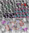
Figure 2. Electron density map of phosphorylated smHMM
The color scheme for all atomic models, unless otherwise noted, is as follows: the HC of the free-like myosin head is magenta, and the HC of the blocked-like head is red. For both myosin heads, the converter domain is green, the ELC blue, and the RLC orange. (A) Averaged in-plane projection of frozen hydrated arrays with a surface view of the 3D reconstruction as Inset. Outlined are two possible fits of the phosphorylated smHMM model: I (magenta) intermolecular head-to-head contact; II (green) intramolecular head-to-head contact. The view orientation is perpendicular to the plane of the crystal. The lipid monolayer would be located below the structure in this view. (B) View of the open smHMM atomic model in space filling, and dimers of the open intermolecular head-to-head contact model (shown in ribbon view in light blue and magenta). (C) A pair of models from the best fit of the open configuration from MDERsca-2hep smHMM. Same coloring scheme used here as for the space filling model shown in (B). Contour threshold set at 140 (arbitrary units).
Figure 3
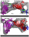
Figure 3. Comparison of the fitting of the closed conformation model with the open conformation model
Adjacent molecules of the same model are also shown. (A) The fit using NMFF of the dephosphorylated closed conformation . A single smHMM molecule is outlined. Two regions of density that are not fit by any feature of the model are marked with asterisks (*). There is no density in the map that can be assigned to an S2 domain for this model. (B) Similar display of the best open conformation model with adjacent molecules shown and one molecule outlined. This interpretation assigns all significant densities to at least one model feature and has a density that can be assigned to S2. This interpretation also has better crystal contacts.
Figure 4
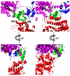
Figure 4. Comparison of the head-to-head interactions of both phosphorylated and dephosphorylated smHMM
(A) View perpendicular to the crystal plane showing the intermolecular interaction between phosphorylated smHMM molecules. The free-like head sequences contributing to the interaction include residues 742-750 (at the end of a disordered loop) and blocked-like head residues contributing include 371-373 and 384-390. (B) The interaction of the dephosphorylated smHMM molecule in a view similar to (A) shows intramolecular binding, where the blocked head binds intimately into the area between the converter and ELC of the free head . (C) View parallel to the crystal plane showing the intermolecular interaction of phosphorylated smHMM. (D) The interaction of the dephosphorylated smHMM molecule in a view similar to (C) shows intramolecular binding, where the blocked head MD binds intimately into the area between the converter and ELC of the free head . The potential areas of interaction for the phosphorylated heads are shown as spheres centered on the Cα atom for clarity.
Figure 5
Figure 5. Phosphorylated and dephosphorylated smHMM oriented as if resting vertically on the lipid monolayer
All views show molecular models fitted to reconstructions (gray) viewed within the crystal plane. The rows of “clothes pins” represent the position of the lipid monolayer. (A) The model of two phosphorylated smHMM molecules from one unit cell shown with their head contacting intermolecularly with the lipid monolayer shown schematically. Direction of view is almost left to right in Figure 2 but along the plane of the array. (B) View of the Wendt et al. model of dephosphorylated smHMM, with the heads bound intramolecularly. The interaction surface in this case is larger than for phosphorylated smHMM. Depth of displayed map is ~85Å.
Figure 6
Figure 6. Comparison of various smooth and scallop muscle LCD models
(A) Side views and end views after rotation by 90° of the HC component of LCD models from the blocked heads of various smooth muscle reconstructions aligned at residues 823-835, or to the corresponding residues in the case of the scallop LCD. (B) Corresponding views for the free heads. The scallop LCD ±Ca2+ is also shown for comparison. The color scheme for HC diagrams is as follows: the blocked heads from the dephosphorylated smHMM models of ; and tarantula are red, the HCs of the corresponding free heads are magenta; the free-like head (this work) is blue, the blocked-like head is yellow; in (A) the HC of the initial MDERsca homology model is cyan and the HC of the MDERske initial model is light green; in (B) the HC of the scallop LCD in low [Ca2+] is gray and in high [Ca2+] is black.
Figure 7
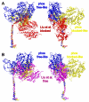
Figure 7. Comparison of phosphorylated and dephosphorylated HCs
All HCs are aligned to residues 780-835 (LCD) of their blocked and blocked-like heads (A) or free and free-like heads (B). Coloring scheme has the free head of the closed, dephosphorylated smHMM - magenta, blocked head - red, free-like head of phosphorylated smHMM – blue, blocked-like head – yellow. Dephosphorylated smHMM atomic model from Liu et al. while the phosphorylated heads are from the MDERsca-2hep model. The diagram shows that the free head of dephosphorylated smHMM is much closer in structure to the free-like head of phosphorylated smHMM than is the blocked head.

References

    1. Sellers JR, Sheterline P, editors. Protein Profile. 2 edit Oxford University Press; Oxford: 1999. Myosins.
    1. Geeves MA, Holmes KC. Structural mechanism of muscle contraction. Annu Rev Biochem. 1999;68:687–728. - PubMed
    1. Trybus KM. Regulation of smooth muscle myosin. Cell Motil Cytoskeleton. 1991;18:81–5. - PubMed
    1. Sellers JR. Mechanism of the phosphorylation-dependent regulation of smooth muscle heavy meromyosin. J Biol Chem. 1985;260:15815–9. - PubMed
    1. Ikebe M, Hartshorne DJ. The role of myosin phosphorylation in the contraction-relaxation cycle of smooth muscle. Experientia. 1985;41:1006–10. - PubMed

Publication types

Substances

Associated data

LinkOut - more resources