

Minor histocompatibility antigens are expressed in syncytiotrophoblast and trophoblast debris: implications for maternal alloreactivity to the fetus
- PMID: 22079431
- PMCID: PMC3338347
- DOI: 10.1016/j.ajpath.2011.09.021
Minor histocompatibility antigens are expressed in syncytiotrophoblast and trophoblast debris: implications for maternal alloreactivity to the fetus
Abstract
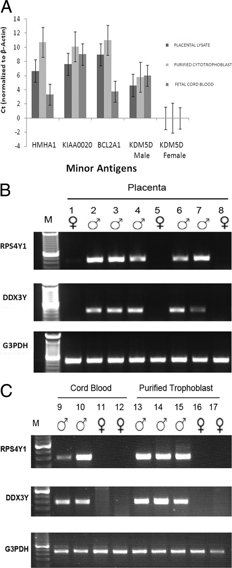
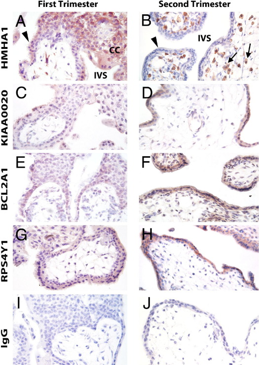
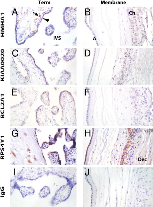
The fetal semi-allograft can induce expansion and tolerance of antigen-specific maternal T and B cells through paternally inherited major histocompatibility complex and minor histocompatibility antigens (mHAgs). The effects of these antigens have important consequences on the maternal immune system both during and long after pregnancy. Herein, we investigate the possibility that the placental syncytiotrophoblast and deported trophoblastic debris serve as sources of fetal mHAgs. We mapped the expression of four mHAgs (human mHAg 1, pumilio domain-containing protein KIAA0020, B-cell lymphoma 2-related protein A1, and ribosomal protein S4, Y linked) in the placenta. Each of these proteins was expressed in several placental cell types, including the syncytiotrophoblast. These antigens and two additional Y chromosome-encoded antigens [DEAD box polypeptide 3, Y linked (DDX3Y), and lysine demethylase5D] were also identified by RT-PCR in the placenta, purified trophoblast cells, and cord blood cells. Finally, we used a proteomic approach to investigate the presence of mHAgs in the syncytiotrophoblast and trophoblast debris shed from first-trimester placenta. By this method, four antigens (DDX3Y; ribosomal protein S4, Y linked; solute carrier 1A5; and signal sequence receptor 1) were found in the syncytiotrophoblast, and one antigen (DDX3Y) was found in shed trophoblast debris. The finding of mHAgs in the placenta and in trophoblast debris provides the first direct evidence that fetal antigens are present in debris shed from the human placenta. The data, thus, suggest a mechanism by which the maternal immune system is exposed to fetal alloantigens, possibly explaining the relationship between parity and graft-versus-host disease.
Copyright © 2012 American Society for Investigative Pathology. Published by Elsevier Inc. All rights reserved.
Figures
Similar articles
-
HLA Class I protein expression in the human placenta.Early Pregnancy (Cherry Hill). 2001 Jan;5(1):67-9. Early Pregnancy (Cherry Hill). 2001. PMID: 11753519
-
Trophoblast expression of the minor histocompatibility antigen HA-1 is regulated by oxygen and is increased in placentas from preeclamptic women.Placenta. 2015 Aug;36(8):832-8. doi: 10.1016/j.placenta.2015.05.018. Epub 2015 Jun 6. Placenta. 2015. PMID: 26095815 Free PMC article.
-
Trophoblast debris modulates the expression of immune proteins in macrophages: a key to maternal tolerance of the fetal allograft?J Reprod Immunol. 2012 Jun;94(2):131-41. doi: 10.1016/j.jri.2012.03.488. Epub 2012 Apr 27. J Reprod Immunol. 2012. PMID: 22542910
-
Fetal tolerance in human pregnancy--a crucial balance between acceptance and limitation of trophoblast invasion.Immunol Lett. 2008 Jan 15;115(1):21-32. doi: 10.1016/j.imlet.2007.09.014. Epub 2007 Nov 5. Immunol Lett. 2008. PMID: 18055021 Review.
-
Maternal and fetal immune responses during pregnancy.Exp Clin Immunogenet. 1993;10(2):85-102. Exp Clin Immunogenet. 1993. PMID: 8251183 Review.
Cited by
-
Trophoblast antigens, fetal blood cell antigens, and the paradox of fetomaternal tolerance.J Exp Med. 2022 May 2;219(5):e20211515. doi: 10.1084/jem.20211515. Epub 2022 Apr 13. J Exp Med. 2022. PMID: 35416936 Free PMC article.
-
Extracellular vesicles and immune response during pregnancy: A balancing act.Immunol Rev. 2022 Jul;308(1):105-122. doi: 10.1111/imr.13074. Epub 2022 Feb 23. Immunol Rev. 2022. PMID: 35199366 Free PMC article. Review.
-
Decidual CD8+T cells exhibit both residency and tolerance signatures modulated by decidual stromal cells.J Transl Med. 2020 Jun 1;18(1):221. doi: 10.1186/s12967-020-02371-3. J Transl Med. 2020. PMID: 32487187 Free PMC article.
-
Specific innate immune cells uptake fetal antigen and display homeostatic phenotypes in the maternal circulation.J Leukoc Biol. 2022 Mar;111(3):519-538. doi: 10.1002/JLB.5HI0321-179RR. Epub 2021 Dec 10. J Leukoc Biol. 2022. PMID: 34889468 Free PMC article.
-
Complex chimerism: pregnancy after solid organ transplantation.Chimerism. 2013 Jul-Sep;4(3):71-7. doi: 10.4161/chim.25401. Epub 2013 Jun 25. Chimerism. 2013. PMID: 23974274 Free PMC article.
References
-
- Petroff M.G. Immune interactions at the maternal-fetal interface. J Reprod Immunol. 2005;68:1–13. - PubMed
-
- Aluvihare V.R., Kallikourdis M., Betz A.G. Regulatory T cells mediate maternal tolerance to the fetus. Nat Immunol. 2004;5:266–271. - PubMed
-
- Darrasse-Jeze G., Klatzmann D., Charlotte F., Salomon B.L., Cohen J.L. CD4+CD25+ regulatory/suppressor T cells prevent allogeneic fetus rejection in mice. Immunol Lett. 2006;102:106–109. - PubMed
-
- Schumacher A., Wafula P.O., Bertoja A.Z., Sollwedel A., Thuere C., Wollenberg I., Yagita H., Volk H.-D., Zenclussen A.C. Mechanisms of action of regulatory T cells specific for paternal antigens during pregnancy. Obstet Gynecol. 2007;110:1137–1145. - PubMed
Publication types
MeSH terms
Substances
Grants and funding
LinkOut - more resources
Full Text Sources
