. 2012 Feb 15;204(1):111-117.
doi: 10.1016/j.jneumeth.2011.10.026. Epub 2011 Nov 4.

Repeated assessment of orthotopic glioma pO(2) by multi-site EPR oximetry: a technique with the potential to guide therapeutic optimization by repeated measurements of oxygen

Affiliations

Repeated assessment of orthotopic glioma pO(2) by multi-site EPR oximetry: a technique with the potential to guide therapeutic optimization by repeated measurements of oxygen

Nadeem Khan et al. J Neurosci Methods. .

Abstract

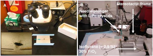
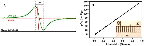
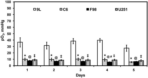
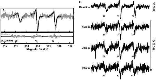
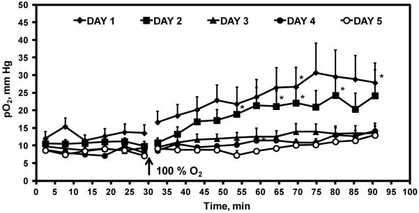
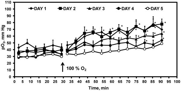
Tumor hypoxia plays a vital role in therapeutic resistance. Consequently, measurements of tumor pO(2) could be used to optimize the outcome of oxygen-dependent therapies, such as, chemoradiation. However, the potential optimizations are restricted by the lack of methods to repeatedly and quantitatively assess tumor pO(2) during therapies, particularly in gliomas. We describe the procedures for repeated measurements of orthotopic glioma pO(2) by multi-site electron paramagnetic resonance (EPR) oximetry. This oximetry approach provides simultaneous measurements of pO(2) at more than one site in the glioma and contralateral cerebral tissue. The pO(2) of intracerebral 9L, C6, F98 and U251 tumors, as well as contralateral brain, were measured repeatedly for five consecutive days. The 9L glioma was well oxygenated with pO(2) of 27-36 mm Hg, while C6, F98 and U251 glioma were hypoxic with pO(2) of 7-12mm Hg. The potential of multi-site EPR oximetry to assess temporal changes in tissue pO(2) was investigated in rats breathing 100% O(2). A significant increase in F98 tumor and contralateral brain pO(2) was observed on day 1 and day 2, however, glioma oxygenation declined on subsequent days. In conclusion, EPR oximetry provides the capability to repeatedly assess temporal changes in orthotopic glioma pO(2). This information could be used to test and optimize the methods being developed to modulate tumor hypoxia. Furthermore, EPR oximetry could be potentially used to enhance the outcome of chemoradiation by scheduling treatments at times of increase in glioma pO(2).


Figures

Figure 1
Figure 1
(A) LiPc crystals are loaded under a microscope in 25 gauge needles (approx 40 - 60 μg) for injection in the rat brain. The insert shows the LiPc crystals, needle and a wooden plunger used to load the crystals in the needle. The bevel of the needles can be blunted to precisely control the depth of the implants and potentially reduce tissue injury. (B) The stereotaxic apparatus used to inject the cells and LiPc aggregates in the brain of rats.
Figure 2
Figure 2
(A) Typical in vitro EPR spectra of the LiPc crystals acquired at 1200 MHz during perfusion with 21% (air) and 0% O2. (B) Change in the line width of the LiPc crystals with change in pO2. This calibration is used to convert the line width observed in vivo to tissue pO2. Each batch of LiPc crystals is calibrated prior to in vivo experiments. The insert shows the size of the LiPc implants (~ 1 mm) used for pO2 measurements by EPR oximetry.
Figure 3
Figure 3
Tissue pO2 of intracerebral 9L, C6, and F98 gliomas in untreated rats. The cells were injected on day -14 and the measurements were started on day 1 for five consecutive days. Mean ± SEM, n = 5 - 6. P < 0.05, *9L vs C6; @9L vs F98; 9L vs U251; #F98 day 1 vs day 4.
Figure 4
Figure 4
Tissue pO2 of the contralateral brain in rats inoculated with 9L, C6, and F98 gliomas on day -14. The pO2 measurements were initiated on day 1 and repeated for four subsequent days by HSR-MS EPR oximetry. Mean ± SEM, n = 5 - 6. P < 0.05, *U251 vs 9L, C6, and F98.
Figure 5
Figure 5
Typical in vivo EPR spectra acquired by HSR-MS EPR oximetry from a rat with an intracerebral F98 tumor in the left hemisphere. (A) The EPR signal on the left was acquired from the LiPc implant in the brain (right hemisphere) and the other two signals were acquired from LiPc implants in the tumor (left-hemisphere). The pO2 reported by the LiPc implants in the tumor were pooled to obtain average tumor pO2. (B) Temporal changes in the pO2 of the contralateral brain and F98 tumor in the rat breathing 30% O2 (baseline) and after the gas was switched to 100% O2.
Figure 6
Figure 6
Temporal changes in intracerebral F98 tumor pO2 in rats breathing 30% O2 (baseline) and after the inhaled gas was switched to 100% O2. The experiment was repeated for five consecutive days. Mean ± SEM, n = 5 - 6. P < 0.05, *pO2 100% O2 vs mean baseline pO2.
Figure 7
Figure 7
Temporal changes in the contralateral brain pO2 of rats breathing 30% O2 (baseline) and when the inhaled gas was switched to 100% O2. The experiment was repeated for five consecutive days. Mean ± SEM, n = 5 - 6. P < 0.05, *pO2 100% O2 vs mean baseline pO2.

References

    1. Ahmad R, Kuppusamy P. Theory, instrumentation, and applications of electron paramagnetic resonance oximetry. Chem Rev. 2010;110:3212–36. - PMC - PubMed
    1. Barth RF, Kaur B. Rat brain tumor models in experimental neuro-oncology: the C6, 9L, T9, RG2, F98, BT4C, RT-2 and CNS-1 gliomas. J Neurooncol. 2009;94:299–312. - PMC - PubMed
    1. Baudelet C, Gallez B. How does blood oxygen level dependent (BOLD) contrast correlate with the oxygen partial pressure (pO2) inside tumors? Mag. Res. Med. 2002;48:980–6. - PubMed
    1. Brurberg KG, Graff BA, Rofstad EK. Temporal heterogeneity in oxygen tension in human melanoma xenografts. British Journal of Cancer. 2003;89:350–6. - PMC - PubMed
    1. Candolfi M, Curtin JF, Nichols WS, Muhammad AG, King GD, Pluhar GE, McNiel EA, Ohlfest JR, Freese AB, Moore PF, Lerner J, Lowenstein PR, Castro MG. Intracranial glioblastoma models in preclinical neuro-oncology: neuropathological characterization and tumor progression. J Neurooncol. 2007;85:133–48. - PMC - PubMed

Publication types

MeSH terms

LinkOut - more resources