. 2012 Feb;142(2):388-98.e1-7.
doi: 10.1053/j.gastro.2011.11.001. Epub 2011 Nov 10.

Restricted heterochromatin formation links NFATc2 repressor activity with growth promotion in pancreatic cancer

Affiliations

Restricted heterochromatin formation links NFATc2 repressor activity with growth promotion in pancreatic cancer

Sandra Baumgart et al. Gastroenterology. 2012 Feb.

Abstract

Background & aims: Transcriptional silencing of the p15(INK4b) tumor suppressor pathway overcomes cellular protection against unrestrained proliferation in cancer. Here we show a novel pathway involving the oncogenic transcription factor nuclear factor of activated T cells (NFAT) c2 targeting a p15(INK4b)-mediated failsafe mechanism to promote pancreatic cancer tumor growth.

Methods: Immunohistochemistry, real-time polymerase chain reaction, immunoblotting, and immunofluorescence microscopy were used for expression studies. Cancer growth was assessed in vitro by [(3)H]thymidine incorporation, colony formation assays, and in vivo using xenograft tumor models. Protein-protein interactions, promoter regulation, and local histone modifications were analyzed by immunoprecipitation, DNA pull-down, reporter, and chromatin immunoprecipitation assays.

Results: Our study uncovered induction of NFATc2 in late-stage pancreatic intraepithelial neoplasia lesions with increased expression in tumor cell nuclei of advanced cancers. In the nucleus, NFATc2 targets the p15(INK4b) promoter for inducible heterochromatin formation and silencing. NFATc2 binding to its cognate promoter site induces stepwise recruitment of the histone methyltransferase Suv39H1, causes local H3K9 trimethylation, and allows docking of heterochromatin protein HP1γ to the repressor complex. Conversely, inactivation of NFATc2 disrupts this repressor complex assembly and local heterochromatin formation, resulting in restoration of p15(INK4b) expression and inhibition of pancreatic cancer growth in vitro and in vivo.

Conclusions: Here we describe a novel mechanism for NFATc2-mediated gene regulation and identify a functional link among its repressor activity, the silencing of the suppressor pathway p15(INK4b), and its pancreatic cancer growth regulatory functions. Thus, we provide evidence that inactivation of oncogenic NFATc2 might be an attractive strategy in treatment of pancreatic cancer.


Figures

Figure 1
Figure 1
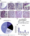
NFATc2 is widely expressed in human pancreatic cancer. (A) Immunohistochemical stainings of NFATc2 in sections of human pancreatic tissues to show that overexpression of NFATc2 is induced in PanIN-2 lesions and is highest in invasively growing cancers. (B) Classification of pancreatic cancers according to NFATc2 expression and localization. (C) Western blot analysis illustrates NFATc2 expression levels in different epithelial pancreatic cancer cell lines. (D) Quantitative real-time PCR to analyze the NFATc2 messenger RNA expression levels in different pancreatic cancer cells. The human cyclophilin gene was used as housekeeping gene control. Highest expression levels of NFATc2 were detected in PaTu8988t and IMIM-PC1 cells.
Figure 2
Figure 2
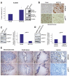
NFATc2 promotes growth of pancreatic cancer. (A and B) Proliferation of (A) PaTu8988t and (B) IMIM-PC1 cells after genetic depletion of NFATc2 in the presence or absence of ionomycin (0.5 μmol/L) was assessed by [3H]thymidine incorporation. Successful knockdown was shown by immunoblotting. (C and D) Suit007 pancreatic cancer cells stably expressing wild-type NFATc2 were subjected to (C) [3H]thymidine incorporation assay or (D) seeded in soft agar. After 10 days, colonies were enumerated. Data are representative of triplicate experiments and displayed ±SD.
Figure 3
Figure 3
NFATc2 blunts p15INK4b expression in vitro and in vivo. (A) Female athymic nude mice were subcutaneously injected with Suit028 cells stably expressing NFATc2 (n = 6) or mock control cells (n = 6). Tumor sizes were measured weekly, and tumor volumes were determined after 5 weeks. Bars represent mean tumor volumes ± SEM. *P = .04. Proteins from xenograft tissues were stained for HA-NFATc2 to prove successful transduction as well as for NFATc2 to confirm overexpression of NFATc2 in Suit028 cells with endogenous low levels of NFATc2. (B) Immunohistochemical analysis to show that NFATc2 and p15INK4b are inversely expressed in Suit028 tumors. (C) Western blot analysis and quantitative real-time PCR to show the reduced p15INK4b expression in NFATc2-overexpressing Suit007 cells. (D) PaTu8988t cells were transfected with NFATc2 siRNA or control siRNA and subjected to Western blot analysis and quantitative real-time PCR to confirm the induction of p15INK4b expression after NFATc2 knockdown. (E) Immunohistochemical analysis of serial sections of human pancreatic tissues to show the inverse expression of NFATc2 and p15INK4b in normal pancreatic tissue, precursor PanIN lesions, and pancreatic cancer.
Figure 4
Figure 4
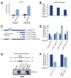
NFATc2 binds to the p15INK4b promoter to mediate its silencing. (A) ChIP analysis using a specific NFATc2 antibody was performed in PaTu8988t cells after 1 hour of treatment with 10% fetal calf serum (FCS). In vivo binding of NFATc2 to the proximal p15INK4b promoter was determined by quantitative real-time PCR using primers specific for the wild-type (wt) p15INK4b promoter region harboring the NFATc2 binding site. (B) PaTu8988t cells were transfected with the wt p15INK4b promoter construct along with increasing amounts of wt NFATc2. Firefly luciferase activities were normalized to Renilla luciferase activity and expressed as fold induction relative to the activity of untreated p15INK4b promoter. (C) Schematic representation of the −751/+75 human full-length p15INK4b promoter construct (wt p15INK4b) and the indicated deletion constructs (p15INK4b del-I-III). The GGAAA NFAT consensus sequence is depicted in the wt p15INK4b promoter construct and also displays the NFAT binding site mutation shown as p15INK4b NFAT-mut. (D) PaTu8988t cells were transfected with the wt p15INK4b or the indicated deletion constructs (p15INK4b del I-III), and responsiveness was measured in the presence or absence of NFATc2. Reporter gene activities are expressed as fold induction related to the control transfected p15INK4b promoter constructs. (E) DNA pull-down experiment to characterize the binding of NFATc2 on the proximal p15INK4b promoter using nuclear extracts from PaTu8988t cells along with either wt p15INK4b or p15INK4b NFAT-mut oligonucleotide sequences. NFATc2 binding was analyzed by immunoblotting. (F) The luciferase activities of the wt p15INK4b and p15INK4b NFAT-mut promoter constructs in PaTu8988t cells were measured in the presence or absence of NFATc2. Reporter gene activities are expressed as fold activation of the p15INK4b promoter constructs relative to the induction without NFATc2.
Figure 5
Figure 5
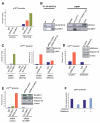
NFATc2 is required for Suv39H1-mediated H3K9 trimethylation and subsequent silencing of the p15INK4b promoter. (A) ChIP analysis was performed in PaTu8988t cells following 1 hour of treatment with 10% fetal calf serum (FCS) using specific NFATc2, H3K9-me, and Suv39H1 antibodies. (B) (Left panel) Coimmunoprecipitation to show the physical interaction between endogenous Suv39H1 and exogenous HA-NFATc2 proteins. (Right panel) DNA pull-down experiment was performed to confirm the requirement of the NFAT binding site for interaction and binding of NFATc2 and Suv39H1 on the p15INK4b promoter. PaTu8988t nuclear lysates were incubated with either wild-type (wt) p15INK4b or p15INK4b NFAT-mut oligonucleotide sequences and subsequently subjected to immunoblotting. (C) Reduced NFATc2, Suv39H1 (left panel), and H3K9-me (right panel) binding to the wt p15INK4b promoter region following transient silencing of NFATc2 in the absence of serum was shown by ChIP assay. Successful knockdown of NFATc2 was confirmed by immunoblotting. H3K9-me values were normalized to native H3. (D and E) Suv39H1-dependent trimethylation of the wt p15INK4b promoter on H3K9 was assessed by ChIP analysis on treatment of PaTu8988t cells with 400 nmol chaetocin to block methyltransferase activity of Suv39H1 (D) or transfection of Suv39H1 siRNA under serum-free conditions (E). Successful treatment and knockdown were evidenced by immunoblotting. (F) IMIM-PC1 cells were transfected with wt p15INK4b promoter along with or without NFATc2 and subjected to luciferase reporter assay following treatment with chaetocin in the absence of serum to show that the NFATc2-mediated silencing of the p15INK4b promoter is dependent on the methyltransferase activity of Suv39H1.
Figure 6
Figure 6
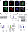
HP1γ stabilizes the repression complex to silence p15INK4b expression. (A) Immunofluorescence staining was performed on transfection of PaTu8988t cells with HA-NFATc2 and GFP-HP1γ constructs and 1 hour of stimulation with 10% fetal calf serum (FCS). Arrows indicate the colocalization of HA-NFATc2 (red signal) and GFP-HP1γ (green signal) in euchromatic foci (weak 4,6-diamidino-2-phenylindole [DAPI] staining within the nucleus) of the nucleus. Nuclei were visualized by DAPI (blue signals) staining. (B) Coimmunoprecipitation to show the Suv39H1 dependency for NFATc2 and HP1γ physical interaction in PaTu8988t cells. (C) PaTu8988t cells were subjected to ChIP analysis after knockdown of Suv39H1 to confirm the requirement of Suv39H1 for HP1γ binding to the wild-type (wt) p15INK4b promoter. (D) DNA pull-down experiment showing the destabilization of NFATc2 and Suv39H1 binding to the wt p15INK4b promoter after knockdown of HP1γ. Successful knockdown is shown by Western blot analysis (lower panel). (E) ChIP analysis was performed to determine HP1γ (left panel) or NFATc2, Suv39H1, and H3K9-me (right panel) binding to the wt p15INK4b promoter following silencing of HP1γ. Successful knockdown was confirmed by Western blotting. (F) Reporter gene assay to prove that the abolishment of HP1γ expression reverses p15INK4b silencing by NFATc2. Reporter gene activities were expressed as fold luciferase activity related to Renilla luciferase activity ±SD.
Figure 7
Figure 7
Pharmacologic targeting of NFATc2 signaling reduces growth of pancreatic cancer and allows re-expression of p15INK4b in vivo. (A and B) IMIM-PC1 cells bearing nude mice were treated intraperitoneally with either 5 mg/kg of the calcineurin inhibitor CsA (n = 5) or 75% dimethyl sulfoxide (DMSO) as a vector control (n = 7). Mice were killed after a treatment period of 2 weeks and tumors were extracted. (A) The mean tumor volume of DMSO- and CsA-treated mice normalized to vector control ±SEM. *P = .01. (B) Messenger RNA was isolated from IMIM-PC1 tumor tissues and quantitative real-time polymerase chain reaction was performed to show the induction of p15INK4b messenger RNA expression following pharmacologic inhibition of NFATc2 shuttling. Mouse 1 was arbitrarily compared with mouse 3 and mouse 2 with mouse 4, respectively.

Similar articles

Cited by

References

    1. Hidalgo M, Maitra A. The hedgehog pathway and pancreatic cancer. N Engl J Med. 2009;361:2094–2096. - PubMed
    1. Singh M, Maitra A. Precursor lesions of pancreatic cancer: molecular pathology and clinical implications. Pancreatology. 2007;7:9–19. - PubMed
    1. Koorstra JB, Hustinx SR, Offerhaus GJ, et al. Pancreatic carcinogenesis. Pancreatology. 2008;8:110–125. - PMC - PubMed
    1. Kim WY, Sharpless NE. The regulation of INK4/ARF in cancer and aging. Cell. 2006;127:265–275. - PubMed
    1. Gil J, Peters G. Regulation of the INK4b-ARF-INK4a tumour suppressor locus: all for one or one for all. Nat Rev Mol Cell Biol. 2006;7:667–677. - PubMed

Publication types

MeSH terms