. 2011 Dec 2;415(4):619-26.
doi: 10.1016/j.bbrc.2011.10.121. Epub 2011 Nov 2.

Tetraspanins regulate the protrusive activities of cell membrane

Affiliations

Tetraspanins regulate the protrusive activities of cell membrane

Rafijul Bari et al. Biochem Biophys Res Commun. .

Abstract

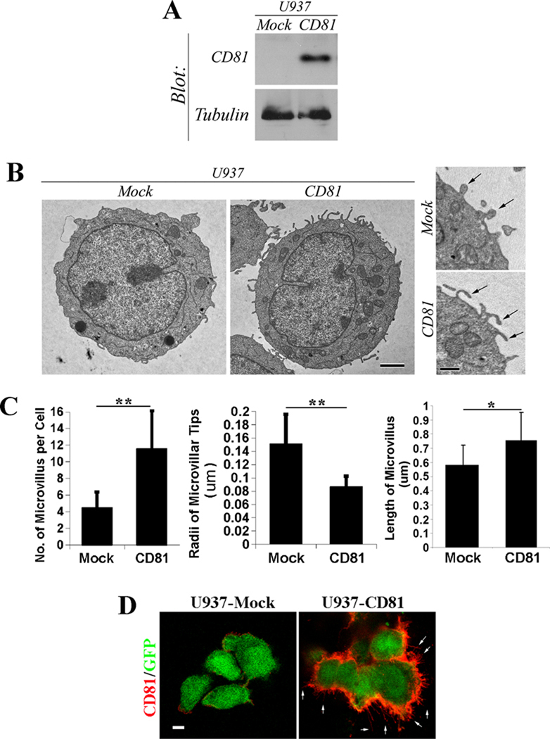
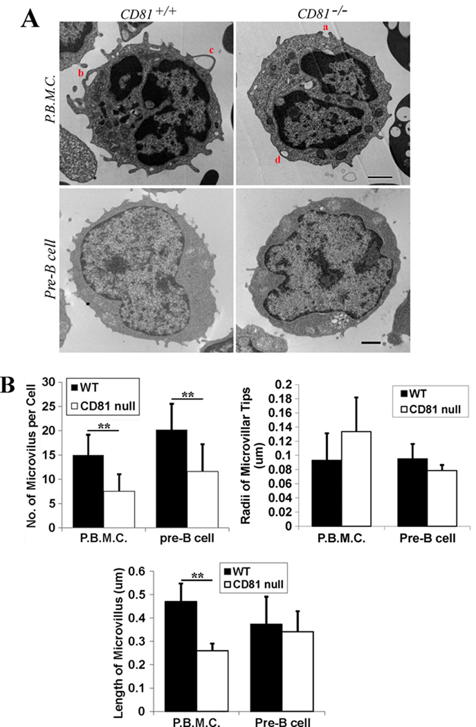
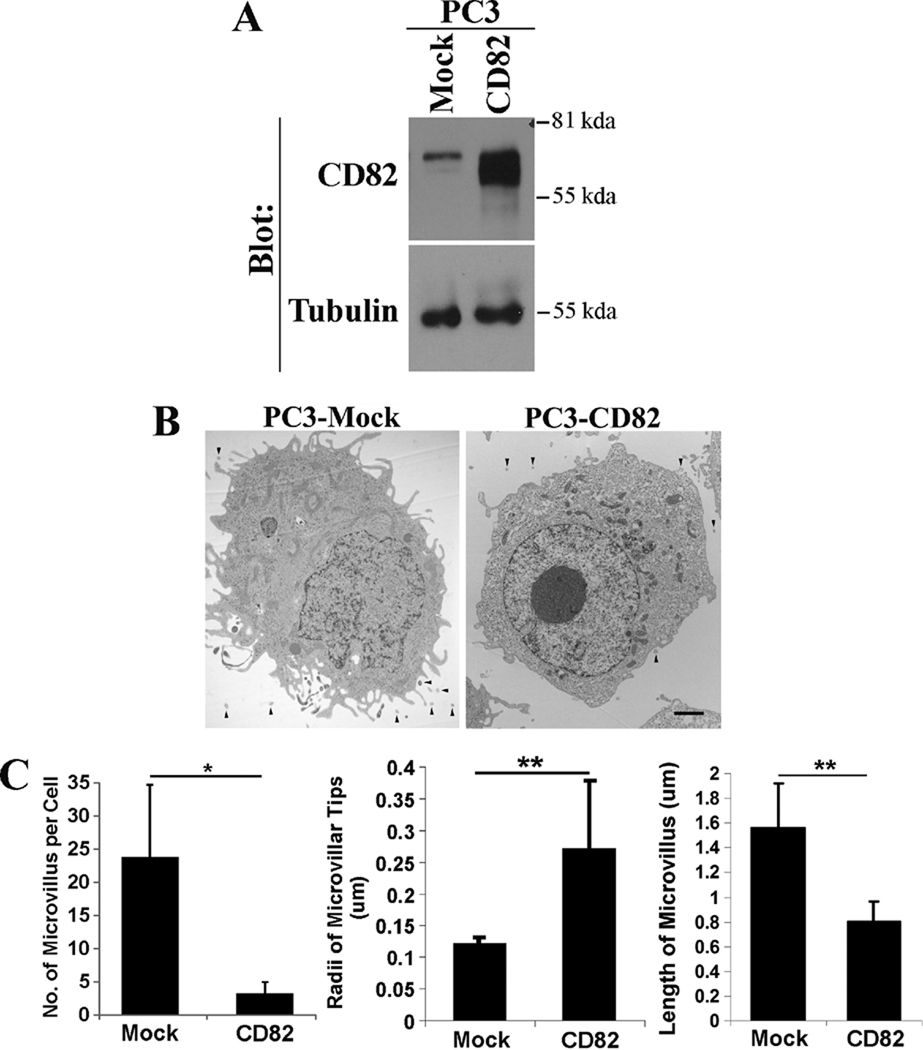
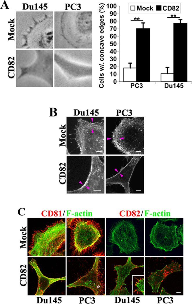
Tetraspanins have gained increased attention due to their functional versatility. But the universal cellular mechanism that governs such versatility remains unknown. Herein we present the evidence that tetraspanins CD81 and CD82 regulate the formation and/or development of cell membrane protrusions. We analyzed the ultrastructure of the cells in which a tetraspanin is either overexpressed or ablated using transmission electron microscopy. The numbers of microvilli on the cell surface were counted, and the radii of microvillar tips and the lengths of microvilli were measured. We found that tetraspanin CD81 promotes the microvillus formation and/or extension while tetraspanin CD82 inhibits these events. In addition, CD81 enhances the outward bending of the plasma membrane while CD82 inhibits it. We also found that CD81 and CD82 proteins are localized at microvilli using immunofluorescence. CD82 regulates microvillus morphogenesis likely by altering the plasma membrane curvature and/or the cortical actin cytoskeletal organization. We predict that membrane protrusions embody a common morphological phenotype and cellular mechanism for, at least some if not all, tetraspanins. The differential effects of tetraspanins on microvilli likely lead to the functional diversification of tetraspanins and appear to correlate with their functional propensity.


Figures

Figure 1
Figure 1. The overexpression of tetraspanin CD81 promotes the formation and extension of microvilli and increases the curvature of microvilli
A: The expressions of CD81 proteins in U937-Mock and -CD81 stable transfectant cells were examined by Western blot. The levels of tubulin proteins are used as loading control. B: The U937 transfectant cells were fixed, thin-sectioned, stained, and analyzed with TEM as described earlier (28). Images represent the results from one out of three individual experiments. Representative images of whole cells and microvilli from the U937 transfectants expressing either control construct or CD81 are presented. Scale bar, 2.0 µm. Arrow: microvillus. C. Quantitative analysis. The numbers of microvilli were counted from individual cells, the radii of microvillar tips were assessed using the transverse cross section of microvilli, and the lengths of microvilli were measured using the longitudinal cross section of microvilli. n=15~17 cells for Mock group and =17~20 cells for CD81 group. *, P<0.05; **, P< or =0.01. D: The localization of CD81 in U937 transfectant cells was examined using immunofluorescence under a confocal microscope. Arrows indicate the microvilli positive in CD81. Scale bar, 5.0 µm.
Figure 2
Figure 2. The ablation of tetraspanin CD81 reduces the formation and probably also extension of microvilli
A: Cells were fixed, thin-sectioned, stained, and analyzed with TEM as described earlier (28). Images represent results from one out of three individual experiments. Representative images of the PBMCs isolated from and pre-B cells derived from the 129/SVJ strain of wild type or CD81-null mice are presented. Scale bar, 0.5 µm for PBMCs or 1.0 µm for pre-B cells. B: Quantitative analysis. The histograms display the mean± SE of 1) the numbers of microvillus per cell, 2) the radii of microvillar tips, and 3) the lengths of microvilli of the PBMCs isolated from wild type or CD81-null mice and the pre-B cell lines derived from wild type (2F3) or CD81-null (1C8) mice (43). For PBMCs, n=12~47 cells for wild type group and =12~39 cells for CD81-null group. For pre-B cells, n=13~45 cells for wild type group and =13~45 cells for CD81-null group. **, P<0.01 between wild type and CD81-null groups.
Figure 3
Figure 3. The overexpression of tetraspanin CD82 inhibits the formation and extension of microvilli
A. The expressions of CD82 proteins in PC3-Mock and -CD82 stable transfectant cells were analyzed with immunoprecipitation followed by immunoblot. Tubulin proteins were examined in Western blot and are used as loading control. B: PC3-Mock and -CD82 cells were fixed, thin-sectioned, stained, and analyzed with TEM as described earlier (28). Scale bar, 2.0 µm. Arrowheads indicate pericellular microvesicles. C. Quantitative analysis. The numbers of microvilli were counted from individual cells, the radii of microvillar tips were assessed using the longitudinal cross section of microvilli, and the lengths of microvilli were measured using the latitudinal cross section of microvilli. n=14 cells for each group. *, P<0.05; **, P<0.01.
Figure 4
Figure 4. Tetraspanin CD82 alters membrane curvature and cortical actin cytoskeleton
A. The cell peripheries become concave upon CD82 expression. Du145-and PC3-Mock and CD82 transfectant cells were spread on fibronectin-coated substratum in serum-free media (for Du145) or cultured in complete media (for PC3). Phase contrast images were obtained under light microscope. CD82-expressing cells constantly formed concave edges, compared with the outward edges, i.e., lamellipodia, in Mock cells. The percentages of the cells with more than one concave edge were quantified. The histograms represent results from 3 experiments (mean ± SD). n=100 cells for each group. **, P<0.01. B. The Du145 and PC3 transfectant cells were fixed, permeabilized, and incubated with Alexa 488-conjugated α-phalloidin. The fluorescent images were acquired with a confocal microscope. Scale bars, 5.0 µm. Arrowheads indicated the cortical actin cytoskeleton. C: The localizations of CD81 and CD82 proteins to microvilli in Du145 and PC3 transfectant cells were examined using immunofluorescence. The cells were fixed, permeabilized, and incubated with Alexa 488-conjugated α-phalloidin and CD81 or CD82 mAb followed by the staining with Alexa 594-conjugated secondary Ab. The images were obtained with confocal microscopy. Scale bar, 5.0 µm. The asterisk indicates the local convex area of a PC3-CD82 transfectant cell.

References

    1. Horejsi V, Vlcek C. Novel structurally distinct family of leucocyte surface glycoproteins including CD9, CD37, CD53 and CD63. FEBS Lett. 1991;288:1–4. - PubMed
    1. Wright MD, Tomlinson MG. The ins and outs of the transmembrane 4 superfamily. Immunol. Today. 1994;15:588–594. - PubMed
    1. Maecker HT, Todd SC, Levy S. The tetraspanin superfamily: molecular facilitators. FASEB J. 1997;11:428–442. - PubMed
    1. Boucheix C, Rubinstein E. Tetraspanins. Cell Mol. Life Sci. 2001;58:1189–1205. - PMC - PubMed
    1. Hemler ME. Tetraspanin functions and associated microdomains. Nat. Rev. Mol. Cell Biol. 2005;6:801–811. - PubMed

Publication types

LinkOut - more resources