Review
. 2012 Apr;69(7):1125-36.
doi: 10.1007/s00018-011-0865-5. Epub 2011 Nov 12.

Microautophagy: lesser-known self-eating

Affiliations
Review

Microautophagy: lesser-known self-eating

Wen-wen Li et al. Cell Mol Life Sci. 2012 Apr.

Abstract

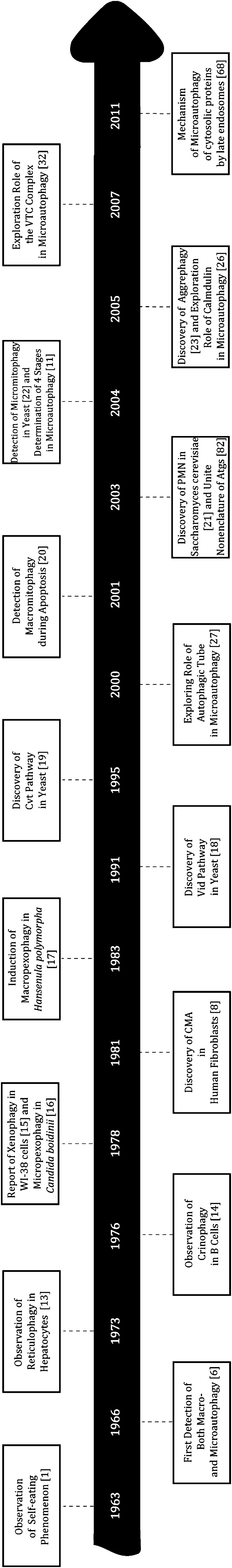
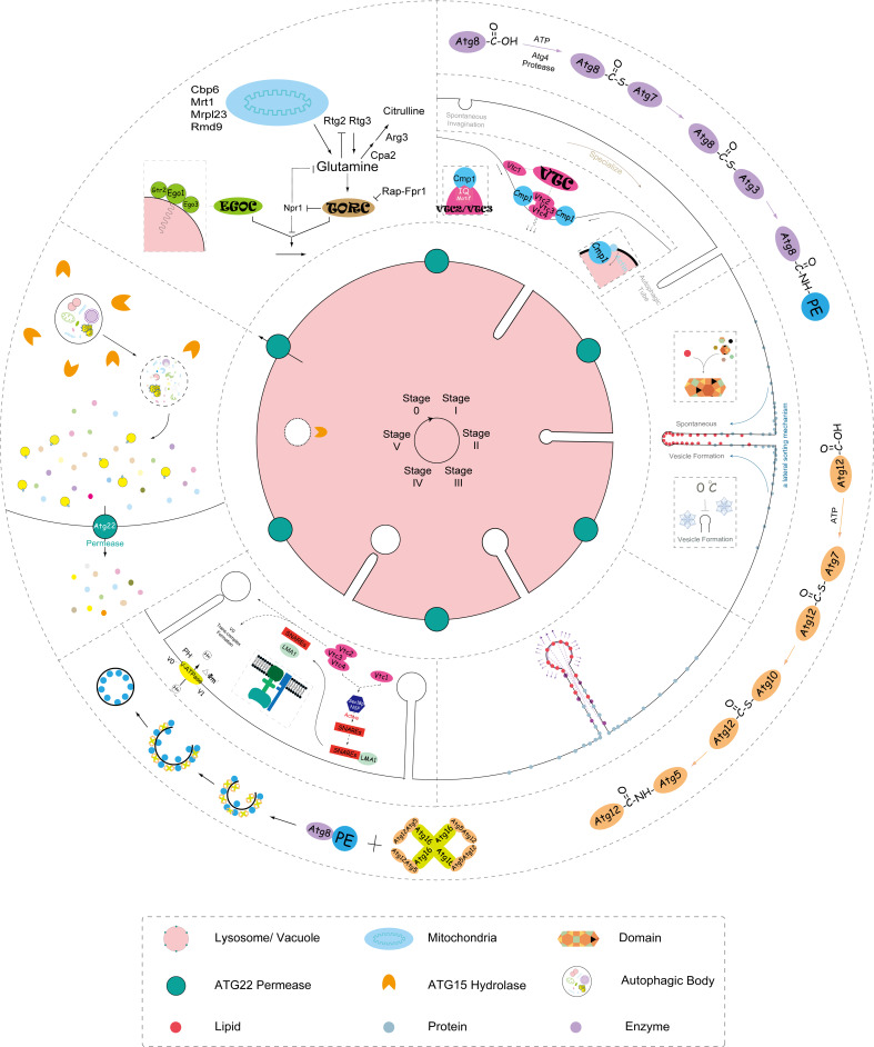
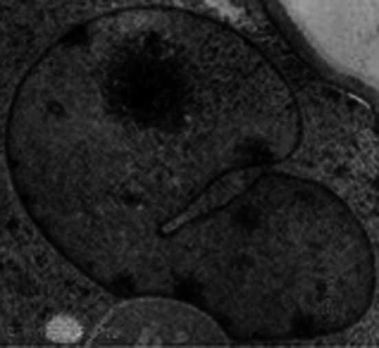
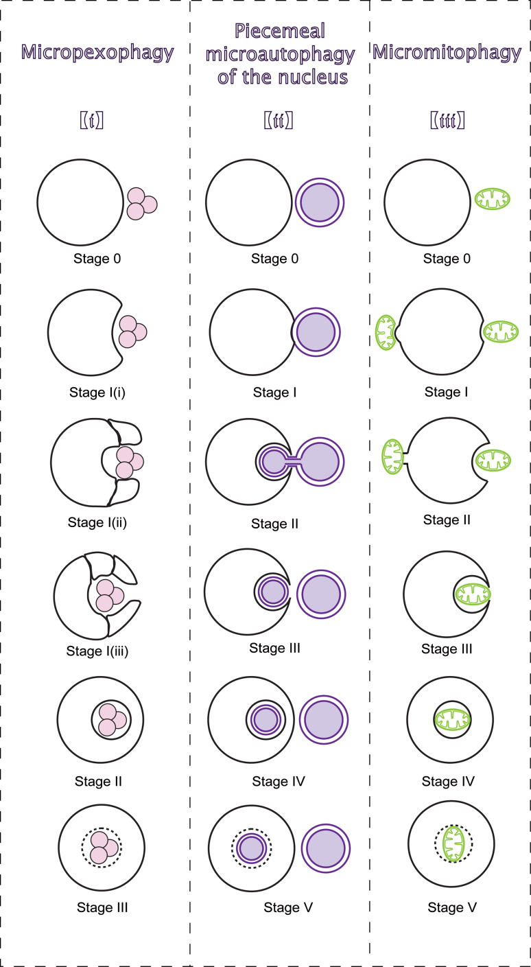
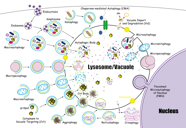
Microautophagy, the non-selective lysosomal degradative process, involves direct engulfment of cytoplasmic cargo at a boundary membrane by autophagic tubes, which mediate both invagination and vesicle scission into the lumen. With its constitutive characteristics, microautophagy of soluble substrates can be induced by nitrogen starvation or rapamycin via regulatory signaling complex pathways. The maintenance of organellar size, membrane homeostasis, and cell survival under nitrogen restriction are the main functions of microautophagy. In addition, microautophagy is coordinated with and complements macroautophagy, chaperone-mediated autophagy, and other self-eating pathways. Three forms of selective microautophagy, including micropexophagy, piecemeal microautophagy of the nucleus, and micromitophagy, share common ground with microautophagy to some degree. As the accumulation of experimental data, the precise mechanisms that govern microautophagy are becoming more appreciated. Here, we review the microautophagic molecular machinery, its physiological functions, and relevance to human diseases, especially in diseases involving multivesicular bodies and multivesicular lysosomes.


Figures

Fig. 1
Fig. 1
Autophagy-related timeline. It presents the time when different forms of self-eating and some molecular events about microautophagy were first reported, from the first observation of autophagy in 1963 to the latest studies of microautophagy by MVBs in 2011
Fig. 2
Fig. 2
Scheme of microautophagy. Every step of microautophagy is presented in a circle on the lysosome/vacuole, including the signaling complexes regulating (e.g., TOR and EGO), membrane invagination, autophagic tubes formation, vesicle formation, vesicle expansion, vesicle scission, degradation of autophagic body, and energy and nutrient recycling
Fig. 3
Fig. 3
The autophagic tube mediates microautophagy, generating a constriction at the neck of the tube, which is what distinguishes autophagic tube from ordinary invagination
Fig. 4
Fig. 4
Morphological sequences of selective microautophagy. The figure divides three types of selective microautophagy into several sequential stages. (i) Micropexophagy. Juxtaposed next to peroxisomes, vacuole receives signals and protrudes VSM along the peroxisome surface. MIPA mediates fusion with the VSM to degrade peroxisomes. (ii) PMN. Though NV junctions, nuclear ER hollows into lysosomal/vacuolar membrane invagination. Piecemeal nucleus is released into the lumen and degraded after fission from the ER. (iii) Micromitophagy. In type I micromitophagy, lysosome/vacuole selectively sequesters mitochondria for degradation. In type II micromitophagy, the vacuole receives signals from the mitochondrion and continuously gets closer to the mitochondrion. Then, mitochondrion heaves at the mitochondria–membrane border, and connects to the vacuole for degradation
Fig. 5
Fig. 5
Fourteen forms of lysosomophagy. The morphological steps of 14 forms of lysosomophagy are elaborated, including macroautophagy, microautophagy, CMA, macropexophagy, macromitophagy, reticulophagy, crinophagy, xenophagy, aggrephagy, micropexophagy, micromitophagy, PMN, Cvt, and Vid

References

    1. deDuve C. The lysosome. Sci Am. 1963;208:64–72. doi: 10.1038/scientificamerican0563-64. - DOI - PubMed
    1. Klionsky DJ. Autophagy revisited: a conversation with Christian de Duve. Autophagy. 2008;4:740–743. - PubMed
    1. Cecconi F, Levine B. The role of autophagy in mammalian development: cell makeover rather than cell death. Dev Cell. 2008;15:344–357. doi: 10.1016/j.devcel.2008.08.012. - DOI - PMC - PubMed
    1. Dalby KN, Tekedereli I, Lopez-Berestein G, Ozpolat B. Targeting the prodeath and prosurvival functions of autophagy as novel therapeutic strategies in cancer. Autophagy. 2010;6:322–329. doi: 10.4161/auto.6.3.11625. - DOI - PMC - PubMed
    1. Kroemer G, Levine B. Autophagic cell death: the story of a misnomer. Nat Rev Mol Cell Bio. 2008;9:1004–1010. doi: 10.1038/nrm2529. - DOI - PMC - PubMed

Publication types