. 2012 Jun;61(6):817-26.
doi: 10.1007/s00262-011-1140-1. Epub 2011 Nov 12.

Enhanced cytotoxicity and decreased CD8 dependence of human cancer-specific cytotoxic T lymphocytes after vaccination with low peptide dose

Affiliations

Enhanced cytotoxicity and decreased CD8 dependence of human cancer-specific cytotoxic T lymphocytes after vaccination with low peptide dose

Tanja Lövgren et al. Cancer Immunol Immunother. 2012 Jun.

Abstract

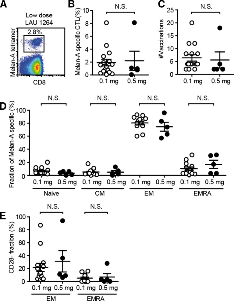
In mice, vaccination with high peptide doses generates higher frequencies of specific CD8+ T cells, but with lower avidity compared to vaccination with lower peptide doses. To investigate the impact of peptide dose on CD8+ T cell responses in humans, melanoma patients were vaccinated with 0.1 or 0.5 mg Melan-A/MART-1 peptide, mixed with CpG 7909 and Incomplete Freund's adjuvant. Neither the kinetics nor the amplitude of the Melan-A-specific CD8+ T cell responses differed between the two vaccination groups. Also, CD8+ T cell differentiation and cytokine production ex vivo were similar in the two groups. Interestingly, after low peptide dose vaccination, Melan-A-specific CD8+ T cells showed enhanced degranulation upon peptide stimulation, as assessed by CD107a upregulation and perforin release ex vivo. In accordance, CD8+ T cell clones derived from low peptide dose-vaccinated patients showed significantly increased degranulation and stronger cytotoxicity. In parallel, Melan-A-specific CD8+ T cells and clones from low peptide dose-vaccinated patients expressed lower CD8 levels, despite similar or even stronger binding to tetramers. Furthermore, CD8+ T cell clones from low peptide dose-vaccinated patients bound CD8 binding-deficient tetramers more efficiently, suggesting that they may express higher affinity TCRs. We conclude that low peptide dose vaccination generated CD8+ T cell responses with stronger cytotoxicity and lower CD8 dependence.


Figures

Fig. 1
Fig. 1
Similar frequencies and differentiation of Melan-A-specific CD8+ T cells after vaccination with low and high peptide doses. Percentages of circulating Melan-A-specific cells in CD8+ T cells after vaccination with 0.1 mg peptide (low dose; n = 16 patients) and 0.5 mg peptide (high dose; n = 5) determined directly ex vivo by flow cytometry. Shown are a representative dot plot of ex vivo Melan-A-specific CD8+ T cells in patient blood after four vaccinations (a), percentages of Melan-A-specific T cells at time of peak response (b), the number of vaccinations needed to reach this peak (c) and the differentiation status of the specific CD8+ T cells, determined as fraction of naïve (CCR7+ CD45RA+), central memory (CM; CCR7+ CD45RA−), effector memory (EM; CCR7−, CD45RA−) and effector RA+ (EMRA; CCR7−, CD45RA+) cells within Melan-A-specific CD8+ T cells (d) and fraction of CD28-cells (e) in Melan-A-specific EM or EMRA subpopulations. Data points for each patient and mean ± SEM are shown. N.S. not significant
Fig. 2
Fig. 2
Equal capacity of cytokine production by Melan-A-specific CD8+ T cells from patients vaccinated with low and high doses of Melan-A peptide. CD8+ T cells from low (n = 6) and high (n = 5) dose-vaccinated patients were analyzed directly ex vivo, upon triggering with unpulsed or Melan-A peptide-pulsed T2 cells for 4 h, followed by intracellular staining for TNF-α (a, b) and IL-2 (a, c). Dot plots of Melan-A-specific CD8+ T cells are shown from two representative patients (a) and percentages of cytokine positive cells for each patient, with mean ± SEM (b, c)
Fig. 3
Fig. 3
Superior capacity of degranulation and target cell lysis by Melan-A-specific CD8+ T cells from low peptide dose-vaccinated patients. Melan-A-specific CD8+ clones were assessed for ability to lyse Melan-A peptide-pulsed T2 target cells (a, n = 323 for low, n = 94 for high peptide dose) or melanoma cell lines (b, n = 14 for low, n = 15 for high peptide dose) positive (Me290) or negative (Na8) for Melan-A expression, with (+ELA) and without additional peptide. Percent specific lysis (a) or lytic units within 107 effector cells (b) are shown as individual data points for each clone, and mean ± SEM for the two patient groups. Ability of ex vivo Melan-A-specific CD8+ T cells (ce, n = 6 for low, n = 5 for high peptide dose) or clones (f, n = 22 for low, n = 17 for high peptide dose) to degranulate was measured as surface expression of CD107a during peptide stimulation. Shown are representative histograms for CD107a expression by Melan-A-specific CD8+ T cells analyzed ex vivo (c), after stimulation with T2 cells without or with Melan-A peptide. Percentages of positive cells (d) or expression per cell (e, mean MFI ± SEM) by ex vivo Melan-A-specific CD8+ T cells, or by Melan-A-specific clones (f, mean MFI ± SEM) for each specified group are depicted
Fig. 4
Fig. 4
Similar steady-state expression of perforin/granzyme B, but increased capacity of perforin release by Melan-A-specific CD8+ T cells from patients vaccinated with low peptide dose. Intracellular staining of perforin (a, c, d) and granzyme B (b, e) was performed in ex vivo Melan-A-specific CD8+ T cells (ae, n = 6 for low, n = 5 for high peptide dose) or Melan-A-specific CTL clones (f, g) from vaccinated patients. Depicted are percentages of perforin (a) or granzyme B (b) positive Melan-A-specific CD8+ T cells for the two patient groups, or percentages of specific cells that lost intracellular expression of perforin upon 4 h stimulation with peptide-loaded target cells (c). Results are shown per patient/clone as well as mean ± SEM. Also shown are mean MFI ± SEM for specific cells (d, e) or clones (f, g; n = 43 for low, n = 20 for high peptide dose)
Fig. 5
Fig. 5
Decreased CD8 dependence but similar avidity of Melan-A-specific CD8+ T cells from low peptide dose-vaccinated patients. Functional avidity of Melan-A-specific clones was determined as logEC50 (M) of Melan-A peptide (a). Values are depicted as separate data points and mean ± SEM for the specified group (low peptide dose: n = 247, high peptide dose: n = 60). Ex vivo CD8+ T cells (b, c; n = 6 for low, n = 5 for high peptide dose) or Melan-A-specific CD8+ T cell clones (df; CD8: n = 43 for low, n = 20 for high peptide dose, tetramers: n = 29 for low, n = 20 for high peptide dose) from vaccinated patients were stained with wild-type Melan-A tetramers (b, e) or for CD8 (c, f). Clones were also stained with mutated Melan-A tetramer unable to bind CD8 (d). Mean MFI ± SEM is depicted for clones from each specified group

References

    1. Jaeger E, Bernhard H, Romero P, Ringhoffer M, Arand M, Karbach J, Ilsemann C, Hagedorn M, Knuth A. Generation of cytotoxic T-cell responses with synthetic melanoma-associated peptides in vivo: implications for tumor vaccines with melanoma-associated antigens. Int J Cancer. 1996;66(2):162–169. doi: 10.1002/(SICI)1097-0215(19960410)66:2<162::AID-IJC4>3.0.CO;2-0. - DOI - PubMed
    1. Cormier JN, Salgaller ML, Prevette T, Barracchini KC, Rivoltini L, Restifo NP, Rosenberg SA, Marincola FM. Enhancement of cellular immunity in melanoma patients immunized with a peptide from MART-1/Melan A. Cancer J Sci Am. 1997;3(1):37–44. - PMC - PubMed
    1. Speiser DE, Lienard D, Rufer N, Rubio-Godoy V, Rimoldi D, Lejeune F, Krieg AM, Cerottini JC, Romero P. Rapid and strong human CD8+ T cell responses to vaccination with peptide, IFA, and CpG oligodeoxynucleotide 7909. J Clin Invest. 2005;115(3):739–746. - PMC - PubMed
    1. Speiser DE, Romero P. Molecularly defined vaccines for cancer immunotherapy, and protective T cell immunity. Semin Immunol. 2010;22(3):144–154. doi: 10.1016/j.smim.2010.03.004. - DOI - PubMed
    1. Rizzuto GA, Merghoub T, Hirschhorn-Cymerman D, Liu C, Lesokhin AM, Sahawneh D, Zhong H, Panageas KS, Perales MA, Altan-Bonnet G, Wolchok JD, Houghton AN. Self-antigen-specific CD8+ T cell precursor frequency determines the quality of the antitumor immune response. J Exp Med. 2009;206(4):849–866. doi: 10.1084/jem.20081382. - DOI - PMC - PubMed

Publication types

MeSH terms