

PSCDB: a database for protein structural change upon ligand binding
- PMID: 22080505
- PMCID: PMC3245091
- DOI: 10.1093/nar/gkr966
PSCDB: a database for protein structural change upon ligand binding
Abstract
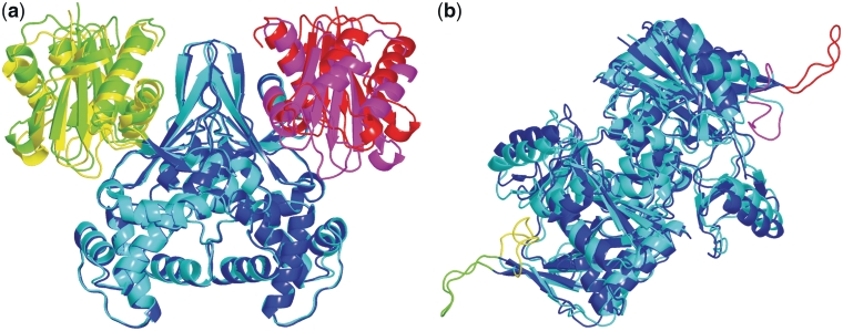
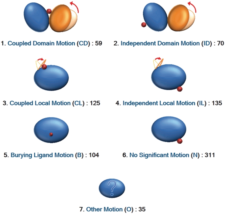
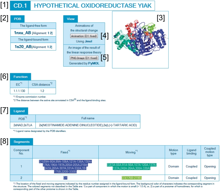
Proteins are flexible molecules that undergo structural changes to function. The Protein Data Bank contains multiple entries for identical proteins determined under different conditions, e.g. with and without a ligand molecule, which provides important information for understanding the structural changes related to protein functions. We gathered 839 protein structural pairs of ligand-free and ligand-bound states from monomeric or homo-dimeric proteins, and constructed the Protein Structural Change DataBase (PSCDB). In the database, we focused on whether the motions were coupled with ligand binding. As a result, the protein structural changes were classified into seven classes, i.e. coupled domain motion (59 structural changes), independent domain motion (70), coupled local motion (125), independent local motion (135), burying ligand motion (104), no significant motion (311) and other type motion (35). PSCDB provides lists of each class. On each entry page, users can view detailed information about the motion, accompanied by a morphing animation of the structural changes. PSCDB is available at http://idp1.force.cs.is.nagoya-u.ac.jp/pscdb/.
Figures
References
-
- Bernstein FC, Koetzle TF, Williams GJ, Meyer EF, Jr, Brice MD, Rodgers JR, Kennard O, Shimanouchi T, Tasumi M. The Protein Data Bank: a computer-based archival file for macromolecular structures. J. Mol. Biol. 1977;112:535–542. - PubMed
-
- Tama F, Sanejouand YH. Conformational change of proteins arising from normal mode calculations. Protein Eng. 2001;14:1–6. - PubMed
-
- Hammes GG. Multiple conformational changes in enzyme catalysis. Biochemistry. 2002;41:8221–8228. - PubMed
-
- Hayward S. Identification of specific interactions that drive ligand-induced closure in five enzymes with classic domain movements. J. Mol. Biol. 2004;339:1001–1021. - PubMed
Publication types
MeSH terms
Substances
LinkOut - more resources
Full Text Sources
Research Materials
