

Role of medullary blood flow in the pathogenesis of renal ischemia-reperfusion injury
- PMID: 22080855
- PMCID: PMC3612396
- DOI: 10.1097/MNH.0b013e32834d085a
Role of medullary blood flow in the pathogenesis of renal ischemia-reperfusion injury
Abstract
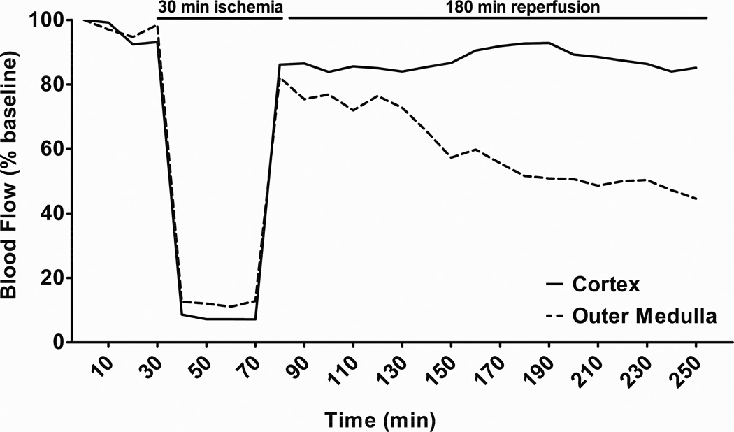
Purpose of review: Renal ischemia-reperfusion injury (IRI) is a common cause of acute kidney injury (AKI). Alterations in renal medullary blood flow (MBF) contribute to the pathogenesis of renal IRI. Here we review recent insights into the mechanisms of altered MBF in the pathogenesis of IRI.
Recent findings: Although cortical blood flow fully recovers following 30-45 min of bilateral IRI, recent studies have indicated that there is a prolonged secondary fall in MBF that is associated with a long-term decline in renal function. Recent findings indicate that angiopoietin-1, atrial natriuretic peptide, heme oxygenase-1, and the gasotransmitters CO and H(2)S, may limit the severity of IRI by preserving MBF. Additional studies have also suggested a role for cytochrome P450-derived 20-HETE in the postischemic fall in MBF.
Summary: Impaired MBF contributes to the pathogenesis of renal IRI. Measurement of renal MBF provides valuable insight into the underlying mechanisms of many renoprotective pathways. Identification of molecules that preserve renal MBF in IRI may lead to new therapies for AKI.
Figures
References
-
- Chertow GM, Burdick E, Honour M, Bonventre JV, et al. Acute kidney injury, mortality, length of stay, and costs in hospitalized patients. J Am Soc Nephrol. 2005;16:3365–3370. - PubMed
-
- Lameire N, Van Biesen W, Vanholder R. The changing epidemiology of acute renal failure. Nat Clin Pract Nephrol. 2006;2:364–377. - PubMed
-
- Lameire N, Vanholder R. Pathophysiologic Features and Prevention of Human and Experimental Acute Tubular Necrosis. J Am Soc Nephrol. 2001;12:S20–S32. - PubMed
-
- Lamiere The changing epidemiology of acute renal failure. Nat Clin Pract Nephrol. 2006;2:364–377. - PubMed
-
- Bonventre JV, Weinberg JM. Recent Advances in the Pathophysiology of Ischemic Acute Renal Failure. J Am Soc Nephrol. 2003;14:2199–2210. - PubMed
Publication types
MeSH terms
Substances
Grants and funding
LinkOut - more resources
Full Text Sources
Research Materials
