. 2011 Dec;121(12):4903-15.
doi: 10.1172/JCI58577. Epub 2011 Nov 14.

Hematopoietic AMPK β1 reduces mouse adipose tissue macrophage inflammation and insulin resistance in obesity

Affiliations

Hematopoietic AMPK β1 reduces mouse adipose tissue macrophage inflammation and insulin resistance in obesity

Sandra Galic et al. J Clin Invest. 2011 Dec.

Abstract

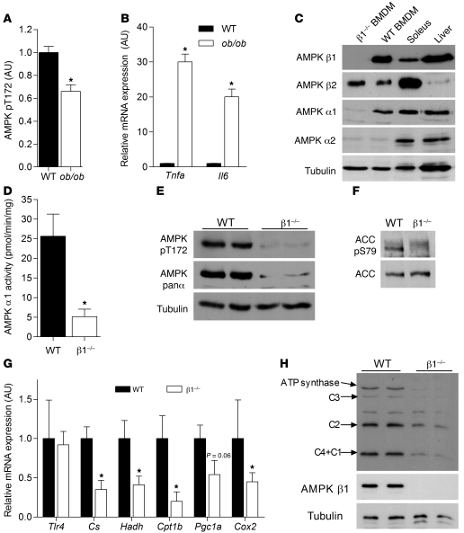
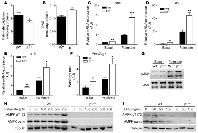
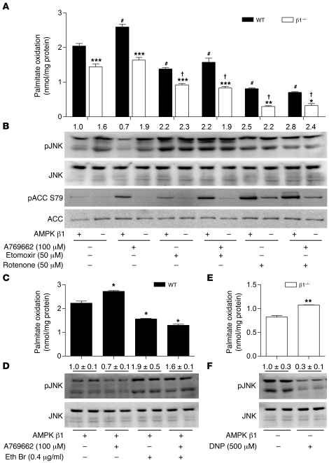
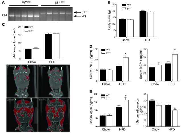
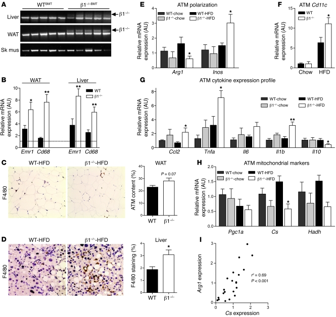
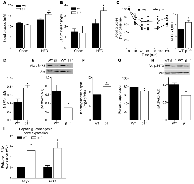
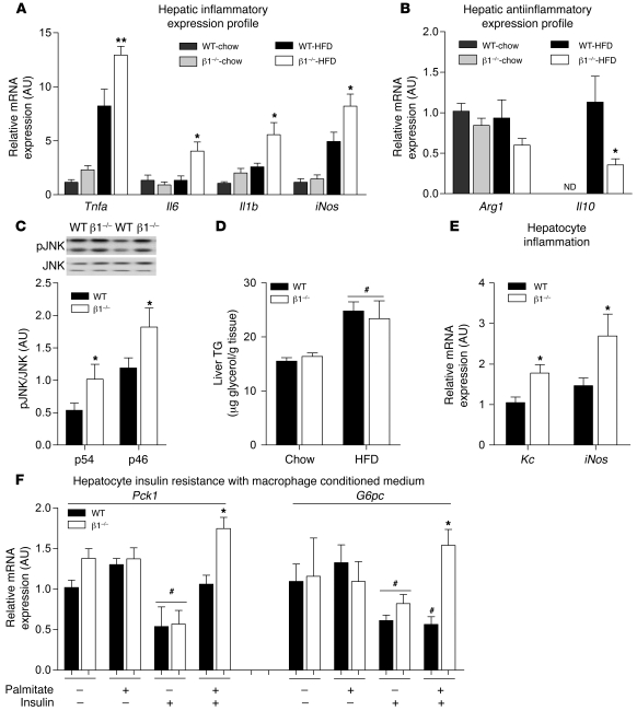
Individuals who are obese are frequently insulin resistant, putting them at increased risk of developing type 2 diabetes and its associated adverse health conditions. The accumulation in adipose tissue of macrophages in an inflammatory state is a hallmark of obesity-induced insulin resistance. Here, we reveal a role for AMPK β1 in protecting macrophages from inflammation under high lipid exposure. Genetic deletion of the AMPK β1 subunit in mice (referred to herein as β1(-/-) mice) reduced macrophage AMPK activity, acetyl-CoA carboxylase phosphorylation, and mitochondrial content, resulting in reduced rates of fatty acid oxidation. β1(-/-) macrophages displayed increased levels of diacylglycerol and markers of inflammation, effects that were reproduced in WT macrophages by inhibiting fatty acid oxidation and, conversely, prevented by pharmacological activation of AMPK β1-containing complexes. The effect of AMPK β1 loss in macrophages was tested in vivo by transplantation of bone marrow from WT or β1(-/-) mice into WT recipients. When challenged with a high-fat diet, mice that received β1(-/-) bone marrow displayed enhanced adipose tissue macrophage inflammation and liver insulin resistance compared with animals that received WT bone marrow. Thus, activation of AMPK β1 and increasing fatty acid oxidation in macrophages may represent a new therapeutic approach for the treatment of insulin resistance.


Figures

Figure 1
Figure 1. Deletion of AMPK β1 from macrophages reduces AMPK α1 protein expression and activity and results in reductions in mitochondrial capacity.
(A) Obese ob/ob mice have reduced AMPK activation, which is associated with increased expression of (B) Tnfa and Il6. Cellular lysates from WT and β1–/– BMDMs were subjected to (C) immunoblot analysis using AMPK α1–, α2–, β1–, and β2–specific antibodies, (D) AMPK α1 activity assays, and (E) immunoblotting for AMPK pT172, AMPK panα, and (F) ACC S79 phosphorylation. (G) mRNA expression of Tlr4 and mitochondrial markers (Cs, Hadh, Cpt1b, Pgc1a, and Cox2 [mitochondrial encoded]) in macrophages from β1–/– and WT mice. (H) Cellular lysates from WT and β1–/– BMDMs were subjected to immunoblot analysis for mitochondrial electron transport chain proteins. C1, complex I subunit 20 kDa; C2, complex II subunit 30 kDa; C3, complex III subunit core 2; C4, complex IV cytochrome oxidase–2. (Note: The unlabeled band is nonspecific, and C4+C1 appeared at approximately the same MW of 20 kDa.) Data are expressed as mean ± SEM; n = 6–8. *P < 0.05 compared with WT, where gene expression was normalized to 18s.
Figure 2
Figure 2. AMPK β1 buffers against lipid accumulation and palmitate-induced macrophage inflammation.
WT and β1–/– BMDMs were incubated with palmitate (0.5 mM, 0.5 μCi/ml 14C-palmitate) for 4 hours and (A) fatty acid oxidation and (B) DAG esterification measured. To investigate the state of macrophage polarization, we incubated WT and β1–/– BMDMs with or without palmitate (0.5 mM) for 24 hours and determined the gene expression of (C) Tnfa, (D) Il6, and (E) Il1b and (F) the ratio of iNos to Arg1 gene expression as a marker of M1/M2 activation. (G) Cells were treated with or without palmitate (0.5 mM) for 4 hours, and phosphorylation of JNK (Thr183/Tyr185) was assessed. (H) WT and β1–/– BMDMs were treated with palmitate at the indicated concentrations for 3 hours and cellular lysates probed with antibodies specific for phosphorylated AMPK T172, total AMPK α protein, and tubulin. (I) WT and β1–/– BMDMs were incubated with increasing concentrations of LPS, and pT172, total AMPK α protein, and tubulin protein expression was determined. Data are expressed as mean ± SEM; n = 3, from at least 2 independent experiments. *P < 0.05, **P < 0.01, ***P < 0.001 compared with WT, where gene expression was normalized to Actb.
Figure 3
Figure 3. Mitochondrial fatty acid oxidation and β1-specific activation of AMPK suppresses JNK phosphorylation in WT but not β1–/– macrophages.
(A) WT and β1–/– BMDMs were treated with 0.5 mM palmitate (0.5 μCi/ml 14C-palmitate) for 4 hours in the presence of A769662 (100 μM), etomoxir (50 μM), and/or rotenone (50 μM) before determination of fatty acid oxidation. (B) BMDMs were incubated as described above with 0.5 mM palmitate and immunoblotted for p-JNK/JNK (T183/Y185) and p-ACC (S79)/ACC as a measure of AMPK activation. Treatment of WT BMDMs with ethidium bromide (Eth Br) (0.4 μg/ml) in the presence or absence of A769662 (100 μM) (C) reduces fatty acid oxidation and increases (D) JNK phosphorylation, blocking the effects of A769662. β1–/– BMDMs were treated with or without dinitrophenol (DNP) (500 μM), and (E) fatty acid oxidation and (F) p-JNK/JNK levels were determined. Data are expressed as mean ± SEM; n = 3–5 from at least 2 independent experiments. *P < 0.05, **P < 0.01, ***P < 0.001 compared with WT; #P < 0.05 compared with WT basal; and P < 0.05 compared with β1–/– basal. For expression of total JNK, duplicate gels were used.
Figure 4
Figure 4. Hematopoietic deletion of AMPK β1 results in systemic inflammation despite similar body mass and adiposity.
WTBMT and β1–/–BMT mice were fed chow or HFD for 22 weeks. (A) PCR analysis of genomic DNA isolated from bone marrow, using WT or β1–/– specific primers. (B) Body weight and (C) total adiposity as determined by CT (with representative captures shown). (D) Serum MCP-1 and TNF-α and (E) serum leptin and adiponectin levels in chow- and HFD-fed mice. Data are expressed as mean ± SEM; n = 8–10. *P < 0.05 compared with WTBMT.
Figure 5
Figure 5. Hematopoietic deletion of AMPK β1 results in macrophage recruitment and inflammatory activation of adipose tissue macrophages.
(A) PCR genotyping of genomic DNA from liver, white adipose tissue (WAT), and skeletal muscle (Sk mus), with WT- or β1-specific primers. (B) mRNA expression of macrophage markers Emr1 (F4/80) and Cd68 in WAT and liver of HFD-fed WTBMT and β1–/–BMT mice (dotted line indicates expression in chow-fed WTBMT mice). Immunohistochemical staining and quantification of F4/80 in (C) WAT and (D) liver (original magnification, ×200). Adipose tissue macro­phage (ATM) mRNA expression of (E) Arg1 and iNos, (F) Itgax (Cd11c), (G) inflammatory cytokines Ccl2, Tnfa, Il6, Il1b, and Il10, and (H) markers of mitochondrial density Pgc1a, Cs, and Hadh. (I) Correlation analysis between expression of Cs and Arg1. All data presented are mean ± SEM; n = 8–10. *P < 0.05, **P < 0.01 compared with WTBMT, within dietary treatment, where the relative expression was normalized to Actb.
Figure 6
Figure 6. Hematopoietic deletion of AMPK β1 causes adipose tissue and hepatic insulin resistance.
WTBMT and β1–/–BMT mice were fed a chow diet or HFD for 22 weeks, and (A) fed blood glucose and (B) serum insulin levels were determined. (C) WTBMT and β1–/–BMT mice fed HFD were fasted for 6 hours and insulin tolerance tests performed after intraperitoneal injection of 0.7 U/kg insulin (right: AUC calculated as mM*min). Hyperinsulinemic-euglycemic clamps were performed in WTBMT and β1–/–BMT fed HFD, and (D) post-clamp serum NEFA and (E) adipose tissue Akt (pS473) phosphorylation were determined. (F) Hepatic glucose output and (G) percent suppression of hepatic glucose production were measured, and livers were collected after the clamp for analysis of (H) phosphorylated Akt (S473) and (I) mRNA expression of gluconeogenic enzymes G6pc and Pck1. All data presented are mean ± SEM; n = 8–10. *P < 0.05 compared with WTBMT within dietary treatment, where relative expression was normalized to Actb. For expression of total Akt, duplicate gels were used.
Figure 7
Figure 7. Increased hepatic inflammation due to β1 deletion in macrophages leads to hepatic insulin resistance.
(A) mRNA expression inflammatory cytokine production in liver tissue from WTBMT and β1–/–BMT mice on chow and HFD: Il1b, Tnfa, Il6, and iNos. (B) Antiinflammatory marker expression in WTBMT and β1–/–BMT livers: Arg1 and Il10. (C) Immunoblotting for phosphorylated JNK (T183/Y185) and total JNK between genotypes fed HFD. (D) Liver triglyceride (TG) levels in WTBMT and β1–/–BMT mice fed chow or HFD. Primary hepatocytes were incubated with conditioned medium from 0.5 mM palmitate–treated WT and β1–/– BMDMs for 4 hours (as described in Methods). (E) The mRNA expression of markers of hepatocyte inflammation Kc and iNos and (F) the mRNA expression of gluconeogenic genes following 4 hours treatment with 10 nM insulin. All data shown are mean ± SEM; n = 8–10 for animal experiments. In vitro work was performed in triplicate from at least 2 livers. *P < 0.05, **P < 0.01 compared with WT, within treatment group; #P < 0.05 between basal and insulin treatment within genotype, where expression was normalized to Actb. For expression of total JNK, duplicate gels were used. ND, not detected.

Comment in

References

    1. Hotamisligil GS, Shargill NS, Spiegelman BM. Adipose expression of tumor necrosis factor-alpha: direct role in obesity-linked insulin resistance. Science. 1993;259(5091):87–91. doi: 10.1126/science.7678183. - DOI - PubMed
    1. Steinberg GR, et al. Tumor necrosis factor alpha-induced skeletal muscle insulin resistance involves suppression of AMP-kinase signaling. Cell Metab. 2006;4(6):465–474. doi: 10.1016/j.cmet.2006.11.005. - DOI - PubMed
    1. Ghanim H, Aljada A, Hofmeyer D, Syed T, Mohanty P, Dandona P. Circulating mononuclear cells in the obese are in a proinflammatory state. Circulation. 2004;110(12):1564–1571. doi: 10.1161/01.CIR.0000142055.53122.FA. - DOI - PubMed
    1. Weisberg SP, McCann D, Desai M, Rosenbaum M, Leibel RL, Ferrante AW., Jr Obesity is associated with macrophage accumulation in adipose tissue. J Clin Invest. 2003;112(12):1796–1808. - PMC - PubMed
    1. Xu H, et al. Chronic inflammation in fat plays a crucial role in the development of obesity-related insulin resistance. J Clin Invest. 2003;112(12):1821–1830. - PMC - PubMed

Publication types

MeSH terms