

Mutant huntingtin, abnormal mitochondrial dynamics, defective axonal transport of mitochondria, and selective synaptic degeneration in Huntington's disease
- PMID: 22080977
- PMCID: PMC3249480
- DOI: 10.1016/j.bbadis.2011.10.016
Mutant huntingtin, abnormal mitochondrial dynamics, defective axonal transport of mitochondria, and selective synaptic degeneration in Huntington's disease
Abstract
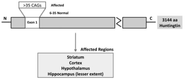
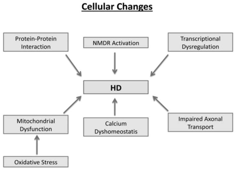
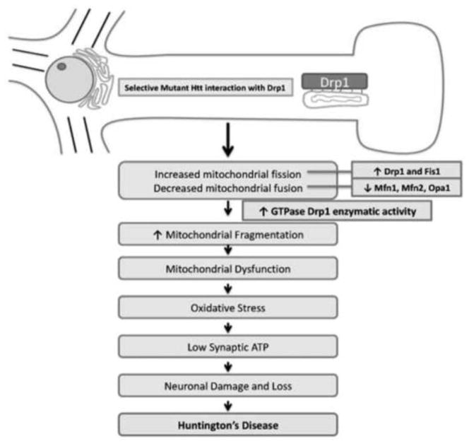
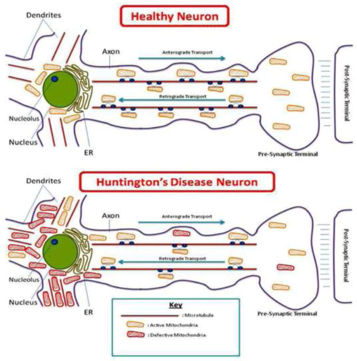
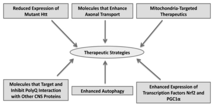
Huntington's disease (HD) is a progressive, fatal neurodegenerative disease caused by expanded polyglutamine repeats in the HD gene. HD is characterized by chorea, seizures, involuntary movements, dystonia, cognitive decline, intellectual impairment and emotional disturbances. Research into mutant huntingtin (Htt) and mitochondria has found that mutant Htt interacts with the mitochondrial protein dynamin-related protein 1 (Drp1), enhances GTPase Drp1 enzymatic activity, and causes excessive mitochondrial fragmentation and abnormal distribution, leading to defective axonal transport of mitochondria and selective synaptic degeneration. This article summarizes latest developments in HD research and focuses on the role of abnormal mitochondrial dynamics and defective axonal transport in HD neurons. This article also discusses the therapeutic strategies that decrease mitochondrial fragmentation and neuronal damage in HD.
Copyright © 2011 Elsevier B.V. All rights reserved.
Figures
Similar articles
-
Mutant huntingtin's interaction with mitochondrial protein Drp1 impairs mitochondrial biogenesis and causes defective axonal transport and synaptic degeneration in Huntington's disease.Hum Mol Genet. 2012 Jan 15;21(2):406-20. doi: 10.1093/hmg/ddr475. Epub 2011 Oct 13. Hum Mol Genet. 2012. PMID: 21997870 Free PMC article.
-
Mitochondrial Abnormalities and Synaptic Damage in Huntington's Disease: a Focus on Defective Mitophagy and Mitochondria-Targeted Therapeutics.Mol Neurobiol. 2021 Dec;58(12):6350-6377. doi: 10.1007/s12035-021-02556-x. Epub 2021 Sep 14. Mol Neurobiol. 2021. PMID: 34519969 Review.
-
Abnormal mitochondrial dynamics, mitochondrial loss and mutant huntingtin oligomers in Huntington's disease: implications for selective neuronal damage.Hum Mol Genet. 2011 Apr 1;20(7):1438-55. doi: 10.1093/hmg/ddr024. Epub 2011 Jan 21. Hum Mol Genet. 2011. PMID: 21257639 Free PMC article.
-
The regulation of autophagosome dynamics by huntingtin and HAP1 is disrupted by expression of mutant huntingtin, leading to defective cargo degradation.J Neurosci. 2014 Jan 22;34(4):1293-305. doi: 10.1523/JNEUROSCI.1870-13.2014. J Neurosci. 2014. PMID: 24453320 Free PMC article.
-
Mechanisms for neuronal cell death and dysfunction in Huntington's disease: pathological cross-talk between the nucleus and the mitochondria?J Mol Med (Berl). 2001 Jul;79(7):375-81. doi: 10.1007/s001090100223. J Mol Med (Berl). 2001. PMID: 11466559 Review.
Cited by
-
Impact of pharmacological agents on mitochondrial function: a growing opportunity?Biochem Soc Trans. 2019 Dec 20;47(6):1757-1772. doi: 10.1042/BST20190280. Biochem Soc Trans. 2019. PMID: 31696924 Free PMC article. Review.
-
Huntington's Disease: Calcium Dyshomeostasis and Pathology Models.Acta Naturae. 2017 Apr-Jun;9(2):34-46. Acta Naturae. 2017. PMID: 28740725 Free PMC article.
-
A new method for quantifying mitochondrial axonal transport.Protein Cell. 2016 Nov;7(11):804-819. doi: 10.1007/s13238-016-0268-3. Epub 2016 May 25. Protein Cell. 2016. PMID: 27225265 Free PMC article.
-
Juvenile Huntington's Disease Skin Fibroblasts Respond with Elevated Parkin Level and Increased Proteasome Activity as a Potential Mechanism to Counterbalance the Pathological Consequences of Mutant Huntingtin Protein.Int J Mol Sci. 2019 Oct 26;20(21):5338. doi: 10.3390/ijms20215338. Int J Mol Sci. 2019. PMID: 31717806 Free PMC article.
-
Inhibition of mitochondrial fragmentation diminishes Huntington's disease-associated neurodegeneration.J Clin Invest. 2013 Dec;123(12):5371-88. doi: 10.1172/JCI70911. Epub 2013 Nov 15. J Clin Invest. 2013. PMID: 24231356 Free PMC article.
References
-
- Vonsattel JP, Myers RH, Stevens TJ, Ferrante RJ, Bird ED, Richardson EP. Neuropathological classification of Huntington’s disease. J Neuropathol Exp Neurol. 1985;44:559–577. - PubMed
-
- Folstein SE. Huntington’s Disease. Johns Hopkins University Press; 1990.
-
- Ross CA, Tabrizi SJ. Huntington’s disease: from molecular pathogenesis to clinical treatment. Lancet Neurol. 2011;10:83–98. - PubMed
-
- Byers RK, Gilles FH, Fung C. Huntington’s disease in children: Neuropathologic study of four cases. Neurology. 1973;23:561–569. - PubMed
Publication types
MeSH terms
Substances
Grants and funding
LinkOut - more resources
Full Text Sources
Other Literature Sources
Medical
Research Materials
Miscellaneous
