. 2011 Nov 13;17(12):1641-5.
doi: 10.1038/nm.2464.

A MEK-independent role for CRAF in mitosis and tumor progression

Affiliations

A MEK-independent role for CRAF in mitosis and tumor progression

Ainhoa Mielgo et al. Nat Med. .

Abstract

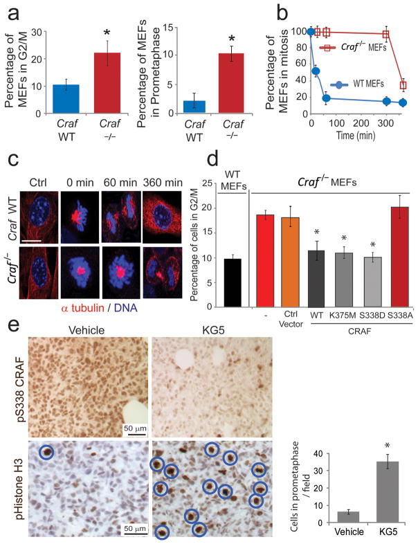
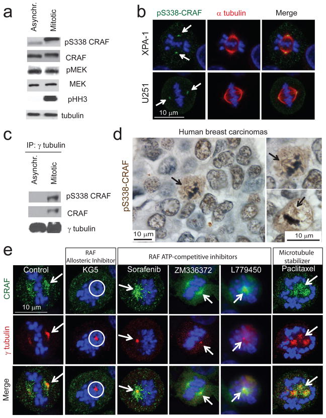
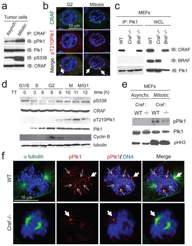
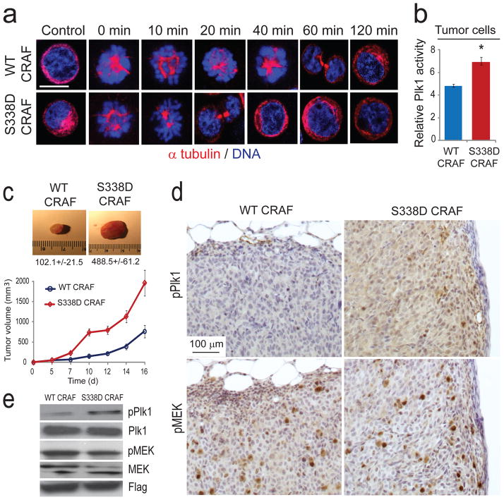
RAF kinases regulate cell proliferation and survival and can be dysregulated in tumors. The role of RAF in cell proliferation has been linked to its ability to activate mitogen-activated protein kinase kinase 1 (MEK) and mitogen-activated protein kinase 1 (ERK). Here we identify a MEK-independent role for RAF in tumor growth. Specifically, in mitotic cells, CRAF becomes phosphorylated on Ser338 and localizes to the mitotic spindle of proliferating tumor cells in vitro as well as in murine tumor models and in biopsies from individuals with cancer. Treatment of tumors with allosteric inhibitors, but not ATP-competitive RAF inhibitors, prevents CRAF phosphorylation on Ser338 and localization to the mitotic spindle and causes cell-cycle arrest at prometaphase. Furthermore, we identify phospho-Ser338 CRAF as a potential biomarker for tumor progression and a surrogate marker for allosteric RAF blockade. Mechanistically, CRAF, but not BRAF, associates with Aurora kinase A (Aurora-A) and Polo-like kinase 1 (Plk1) at the centrosomes and spindle poles during G2/M. Indeed, allosteric or genetic inhibition of phospho-Ser338 CRAF impairs Plk1 activation and accumulation at the kinetochores, causing prometaphase arrest, whereas a phospho-mimetic Ser338D CRAF mutant potentiates Plk1 activation, mitosis and tumor progression in mice. These findings show a previously undefined role for RAF in tumor progression beyond the RAF-MEK-ERK paradigm, opening new avenues for targeting RAF in cancer.


Conflict of interest statement

Competing financial interests

The authors declare no competing financial interests. Published online at http://www.nature.com/naturemedicine/.

Reprints and permissions information is available online at http://npg.nature.com/reprintsandpermissions/.

Figures

Figure 1
Figure 1. CRAF is required for mitotic progression
(a) Cell cycle analysis of WT and Craf−/− MEFs. Left graph, Cells in G2/M were quantified by flow cytometry. Right graph, Cells at pro-metaphase were quantified using confocal microscopy. Error bars represent s.d. (n = 4); * P = 0.0079 (left graph) and P = 0.0055 (right graph) using a Mann Whitney U test. (b) WT and Craf−/− MEFs were synchronized at pro-metaphase with a thymidine-nocodazole block and subsequently released from the blockade and allowed to progress through mitosis. Quantification of cells in G2/M was performed by flow cytometry. Error bars represent s.d. (n = 3). (c) Confocal microscopy images of WT and Craf−/− cells progressing through mitosis at 0, 60 and 360 min after release from pro-metaphase blockade. Cells were stained for α-tubulin (in red) and DNA (TOPRO-3 in blue). Scale bar, 10 μm. (d) WT and Craf−/− MEFs were transfected with vector control, WT CRAF, kinase dead (K375M) CRAF, phospho-mimetic (S338D) CRAF or non-phosphorylatable (S338A) CRAF mutants. Cell cycle analysis and quantification of cells in G2/M was performed by flow cytometry. Error bars represent s.d. (n = 3); * P = 0.0084 using a Mann Whitney U test. (e) Immunohistochemical staining of phospho-S338 CRAF and phospho-histone H3 (mitotic marker) in orthotopic breast and tumor xenografts untreated or treated systemically with 50 mg kg−1 KG5 for 3 d. Scale bar, 50 μm. Circles indicate cells in pro-metaphase. Right, Quantification of cells in pro-metaphase. Error bars represent s.d. (n = 12); * P = 0.006 using Student’s t-test.
Figure 2
Figure 2. Phospho-serine 338 CRAF is upregulated in mitosis and localizes to mitotic spindles in human cell lines and tumor biopsies
(a) Immunoblot analysis of human colon carcinoma HCT-116 cells asynchronized and synchronized at pro-metaphase. pS338 refers to phospho-S338 CRAF, pMEK refers to phospho-MEK and pHH3 refers to phospho-histone H3. Data are representative of three independent experiments. (b) Confocal microscopy images of human pancreatic XPA-1 and glioblastoma U251 cells during mitosis, stained for phospho-S338 CRAF (in green), α-tubulin (in red) and DNA (TOPRO-3 in blue). Scale bar, 10 μm. White arrows indicate localization of phospho-S338 CRAF at the mitotic spindle. (c) Immunoblot analysis of γ-tubulin immunoprecipitates from human colon carcinoma HCT-116 cells asynchronized and synchronized at pro-metaphase. Data are representative of three independent experiments. (d) Immunohistochemical staining of phospho-S338 CRAF in tumor biopsies from breast cancer patients. Scale bar, 10 μm. (e) Confocal microscopy images of XPA-1 cells treated with KG5, sorafenib, ZM336372, L779450 or paclitaxel and stained for CRAF (in green), γ-tubulin (in red) and DNA (TOPRO-3 in blue). Scale bar, 10 μm. White arrows indicate localization of CRAF at the spindle pole. White circle indicates the absence of CRAF at the spindle pole.
Figure 3
Figure 3. CRAF interacts with Plk1 and promotes its activation and accumulation to the kinetochores at pro-metaphase
(a) Immunoblot analysis of CRAF immunoprecipitates from HCT-116 asynchronized and synchronized at pro-metaphase cells. Data are representative of three independent experiments. (b) Confocal microscopy images of HCT-116 cells synchronized at G2 and pro-metaphase (as described in Methods) and stained for CRAF (in green), phospho-T210 Plk1 (in red) and DNA (TOPRO-3 in blue). White arrows indicate co-localization of CRAF with phospho-Plk1 at the centrosomes and mitotic spindle poles. (c) Immunoblot analysis of Plk1 immunoprecipitates from WT, Craf−/− and Braf−/− MEFs. Data are representative of three independent experiments. (d) Immunoblot analysis from G1-M, of phospho-S338 CRAF, total CRAF, phospho-T210 Plk1, total Plk1, cyclin B and tubulin of HCT-116 cells synchronized at the G1/S boundary. Cells were synchronized at the G1/S boundary by a double thymidine block as described in Methods. Data are representative of two independent experiments. (e) Immunoblot analysis of asynchronous and mitotic WT and Craf−/− MEFs. (f) Confocal microscopy images of WT and Craf−/− MEFs at pro-metaphase. Cells were stained for α-tubulin (in green), phospho-T210 Plk1 (in red) and DNA (TOPRO-3 in blue). Thick white arrows indicate localization of phospho-Plk1 at the mitotic spindle pole and narrow white arrows indicate localization of phospho-Plk1 at the kinetochores. Scale bar, 10 μm.
Figure 4
Figure 4. Phospho-mimetic CRAF S338D mutation drives tumor growth and activates Plk1 in vivo
(a) HCT-116 human colon carcinoma cells ectopically expressing either WT RAF or S338D mutant CRAF were arrested in pro-metaphase as described in Methods, and subsequently allowed to progress through mitosis. Cells were stained for α-tubulin (in red) and DNA (TOPRO-3 in blue) at 0, 10, 20, 40, 60 and 120 min after release from pro-metaphase blockade and mitotic progression was analyzed by confocal microscopy. Scale bar, 10 μm. Data are representative of three independent experiments. (b) Plk1 kinase activity assay performed in HCT-116 cells expressing WT CRAF or a phospho-mimetic S338D CRAF mutant. Error bars represent s.d. (n = 3); * P = 0.011 using a Mann Whitney U test. (c) HCT-116 cells expressing WT or S338D CRAF Flag tagged were injected subcutaneously in the flank of immune-compromised nude mice. Tumor images, average weights ± s.e. and tumor size measurements are shown (n = 20). (d) Immunohistochemical staining of phospho-Plk1 and phospho-MEK in mouse tissues from tumors expressing WT or S338D CRAF. Scale bar, 100 μm. (e) Immunoblot analysis of phospho-T210 Plk1, Plk1, phospho-MEK, MEK and FLAG in tumor lysates from HCT-116 cells expressing WT or S338D CRAF. Data are representative of five independent experiments.

References

    1. Wellbrock C, Karasarides M, Marais R. The RAF proteins take centre stage. Nat Rev Mol Cell Biol. 2004;5:875–885. - PubMed
    1. Dhillon AS, Hagan S, Rath O, Kolch W. MAP kinase signalling pathways in cancer. Oncogene. 2007;26:3279–3290. - PubMed
    1. Bollag G, et al. Clinical efficacy of a RAF inhibitor needs broad target blockade in BRAF-mutant melanoma. Nature. 2010;467:596–599. - PMC - PubMed
    1. Flaherty KT, et al. Inhibition of mutated, activated BRAF in metastatic melanoma. N Engl J Med. 2010;363:809–819. - PMC - PubMed
    1. Joseph EW, et al. The RAF inhibitor PLX4032 inhibits ERK signaling and tumor cell proliferation in a V600E BRAF-selective manner. Proc Natl Acad Sci U S A. 2010;107:14903–14908. - PMC - PubMed

Publication types

MeSH terms