. 2011 Nov 13;17(12):1610-8.
doi: 10.1038/nm.2506.

Oxidation of CaMKII determines the cardiotoxic effects of aldosterone

Affiliations

Oxidation of CaMKII determines the cardiotoxic effects of aldosterone

B Julie He et al. Nat Med. .

Abstract

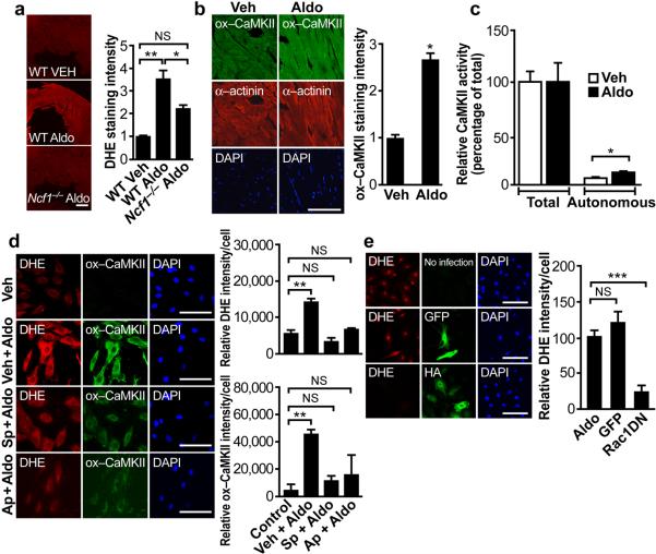
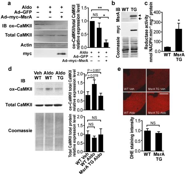
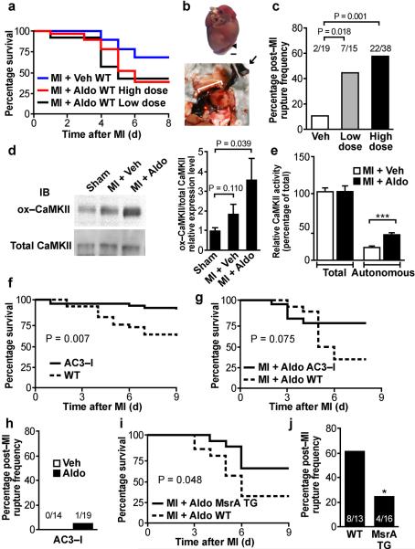
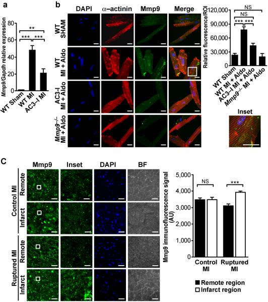
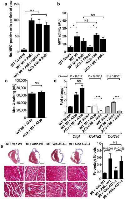
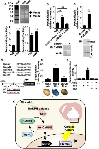
Excessive activation of the β-adrenergic, angiotensin II (Ang II) and aldosterone signaling pathways promotes mortality after myocardial infarction, and antagonists targeting these pathways are core therapies for treating this condition. Catecholamines and Ang II activate the multifunctional Ca(2+)/calmodulin-dependent protein kinase II (CaMKII), the inhibition of which prevents isoproterenol-mediated and Ang II-mediated cardiomyopathy. Here we show that aldosterone exerts direct toxic actions on myocardium by oxidative activation of CaMKII, causing cardiac rupture and increased mortality in mice after myocardial infarction. Aldosterone induces CaMKII oxidation by recruiting NADPH oxidase, and this oxidized and activated CaMKII promotes matrix metalloproteinase 9 (MMP9) expression in cardiomyocytes. Myocardial CaMKII inhibition, overexpression of methionine sulfoxide reductase A (an enzyme that reduces oxidized CaMKII) or NADPH oxidase deficiency prevented aldosterone-enhanced cardiac rupture after myocardial infarction. These findings show that oxidized myocardial CaMKII mediates the cardiotoxic effects of aldosterone on the cardiac matrix and establish CaMKII as a nodal signal for the neurohumoral pathways associated with poor outcomes after myocardial infarction.


Figures

Figure 1
Figure 1
ROS and CaMKII oxidation and activation by Aldo. (a) Representative images of DHE fluorescence in viable, cryopreserved myocardium after 15 minutes Aldo (10−7 mol L−1) stimulation in Ncf1−/− and WT mice. Scale bar = 0.5 mm. Summary data, P = 0.003, One-way ANOVA, *P < 0.05 and **P < 0.01 Bonferroni's multiple comparison test. n ≥ 3 mice per genotype. (b) Representative immunofluorescence images after chronic Aldo infusion (1.44 mg kg−1 d−1, 2 weeks) showing enhanced CaMKII oxidation (ox-CaMKII, green) in left ventricular sections. α-actinin (red) marks myocardium. DAPI (blue) marks nuclei. Scale bar = 100 μm. *P = 0.008. n ≥ 3 mice per treatment. (c) Autonomous, Ca2+/calmodulin-independent and total, Ca2+/calmodulin-dependent CaMKII activity after Aldo and Veh infusion. *P = 0.019, Student's t-test. n ≥ 5 mice per treatment. (d) Aldo treatment for 15 minutes (10−7 mol L−1) increased DHE fluorescence and ox-CaMKII more than Veh-treated WT neonatal myocytes. Pretreatment with spironolactone (Sp, MR antagonist, 10−1 μmol L−1) or apocynin (Ap, NADPH oxidase inhibitor, 100 μmol L−1) reduces Aldo-induced DHE and ox-CaMKII. Summary data for both DHE intensity and ox-CaMKII, P < 0.001, One-way ANOVA, ***P < 0.001 Bonferroni's multiple comparison test versus Control. Scale bar = 100 μm. (e) Expression of an HA tagged dominant negative Rac1 mutant (HA) prevented Aldo stimulated increases in DHE fluorescence. P < 0.001, One-way ANOVA, ***P < 0.001 Bonferroni's multiple comparison test versus Aldo. Scale bar = 100 μm.
Figure 2
Figure 2
Transgenic (TG) myocardial MsrA over-expression reduces CaMKII oxidation. (a) Representative immunoblot of CaMKII oxidation in neonatal myocytes. CaMKII oxidation is reduced by pre-infection with an adenoviral construct over-expressing myc tagged human MsrA. Summary data for n = 3 trials, P = 0.008, One-way ANOVA, *P < 0.05, **P < 0.01, Bonferroni's multiple comparison test versus Aldo-treated, non-infected control. (b) Representative immunoblot shows over-expression of MsrA in hearts from transgenic mice. The myc tagged human MsrA transgene (◀) runs higher than endogenous MsrA (←). (c) Cardiac MsrA activity is increased in transgenic mice compared to WT littermates. *P = 0.035, Student's t-test, n ≥ 3 mice per genotype. (d) WT mice infused with Aldo (1.44 mg kg−1 d−1, 2 weeks) show a substantial trend towards increased CaMKII oxidation by Western blotting in total heart homogenates, P = 0.078. MsrA transgenic mice appear resistant to Aldo-induced CaMKII oxidation. Total cardiac CaMKII expression levels remain similar between MsrA transgenic mice and WT control littermates, n = 6 mice per genotype. (e) DHE fluorescence of viable, cryopreserved myocardium after 15 minutes Aldo (10−7 mol L−1) stimulation. Aldo increases DHE fluorescence similarly in MsrA transgenic and WT hearts. Scale bar = 1 mm. n ≥ 3 mice per genotype.
Figure 3
Figure 3
Aldo increases mortality after MI by promoting myocardial rupture. (a) Kaplan-Meier survival curve for WT mice after MI + Veh versus MI + Aldo. n ≥ 15 mice per treatment. (b) Necropsy of a representative MI + Aldo WT mouse. Forceps (→) retracts clotted blood from ruptured heart. Rupture site (◀). Scale bar = 1 mm. (c) Rupture frequency in WT mice after MI + Aldo, at Low dose and High dose (see Supplemental Fig. 4 and Methods), Chi-square test, P = 0.018 and P = 0.001, respectively. (d) Representative immunoblot from post-MI cardiac lysates for ox-CaMKII two weeks after surgery. Summary data for n = 6 mice per treatment. (e) Autonomous and total CaMKII activity in WT MI + Aldo versus MI + Veh mice. ***P < 0.001, Student's t-test, n ≥ 5 mice per treatment. (f) Kaplan-Meier survival curve for WT littermate and AC3-I mice after MI, n ≥ 30 mice per genotype, P = 0.007. (g) Kaplan-Meier survival curve for AC3-I and WT mice after MI + Aldo. n = 15–19 mice per genotype, P = 0.075. (h) Summary data showing rupture protection in AC3-I mice after MI + Aldo or MI + Veh. (i) Kaplan-Meier survival curve for WT littermate and MsrA transgenic (TG) mice after MI + Aldo. n = 13–16 mice per genotype, P = 0.048. (j) Rupture frequency in WT littermate and MsrA TG mice after MI + Aldo. n = 13–16 mice per genotype, *P = 0.047.
Figure 4
Figure 4
Mmp9 levels in mouse and human hearts. (a) Relative Mmp9 mRNA levels 3 days post-MI in WT and AC3-I mice. P < 0.0001, One-way ANOVA, n ≥ 3 mice per treatment, **P < 0.01, ***P < 0.001, Bonferroni's multiple comparison test. (b) Immunofluorescence of isolated adult mouse ventricular myocytes 24 hours after MI shows increased Mmp9 expression in MI + Aldo compared to Sham operated controls and to Mmp9−/− controls. Myocytes from AC3-I mice have lower Mmp9 expression compared to WT myocytes. All images are maximum projections from z-stacks obtained by confocal microscopy. Inset shows Mmp9 expression is cytoplasmic and appears perpendicular to α-actinin staining. Scale bar = 20 μm. Quantification of immunofluorescence. P < 0.0001, One-way ANOVA, ***P < 0.001, Bonferroni's multiple comparison test. At least 10 regions of interest were selected from n = 5–10 cells per treatment. (c) Immunofluorescence for Mmp9 in human myocardium from MI subjects without rupture (Control MI) and with rupture (Ruptured MI). Images are the maximum projection of z-stacks acquired with confocal microscopy. Brightfield (BF) images show cross-sectional myocardial cells in bundles. Inset shows intracellular staining of Mmp9. Quantification of intracellular regions of interest were confined to cardiomyocytes and show an increased speckled and punctate staining pattern for Mmp9 within cardiomyocytes from humans with ruptured MI and no gradient in Mmp9 signal in control MI samples, P < 0.001, One-way ANOVA, n ≥ 5 specimens per group, ***P < 0.001, Bonferroni's multiple comparison test. Scale bar = 25 μm.
Figure 5
Figure 5
Characterization of inflammatory and fibrotic response between AC3-I and WT mice after MI + Aldo. (a) The number of MPO positive cells identified on immunohistochemistry of heart tissue sections is similar between WT and AC3-I mice 24 hours after MI + Aldo and between ruptured and non-ruptured WT MI + Aldo mice. P < 0.001, One-way ANOVA, n ≥ 3 mice per group, ***P < 0.001, Bonferroni's multiple comparison test. (b) MPO activity is similar between WT and AC3-I mice. MPO activity increases after MI in both WT and AC3-I mice. AC3-I mice show no difference to WT mice even after MI + Aldo. P = 0.001, One-way ANOVA, n ≥ 3 mice per group, *P < 0.05, Bonferroni's multiple comparison test. (c) Cumulative Mac-3 staining is similar in AC3-I mice compared to WT mice 3 days after MI + Aldo. n ≥ 3 mice per group. (d) Differential regulation of pro-fibrotic genes after MI + Aldo in WT and AC3-I mice, Ctgf = connective tissue growth factor, Col1a2 = collagen type I alpha 2, Col3a1 = collagen type III alpha 1, n ≥ 4 mice per group, One-way ANOVA, ***P < 0.001, Bonferroni's multiple comparison test. (e) Masson's trichrome staining shows similar increase in fibrosis after MI + Aldo in both WT and AC3-I. Student's t-test versus MI + Veh, n ≥ 3 mice per group. Black scale bar = 2 mm. White scale bar = 50 μm.
Figure 6
Figure 6
CaMKII promotes cardiac Mmp9 expression and activity. (a) Representative gelatin zymogram for Mmp9 and Mmp2 activity in culture supernatant bathing neonatal myocytes, ***P < 0.001, n = 4 assays per treatment. (b) qRT-PCR for Mmp9 in total RNA isolated from neonatal myocytes after 24 hour Aldo treatment with or without CaMKII knockdown by shRNA. Overall P < 0.001, One-way ANOVA, **P < 0.01 Bonferroni's multiple comparison test. Immunoblot verifies CaMKII knockdown. (c) CaMKII over-expression increases Mmp9 mRNA expression compared to control empty virus, **P = 0.002. Immunoblot verifies over-expression of myc-tagged CaMKII. (d) Alignment of the putative MEF2 binding domain from mouse Mmp9 promoter with bona fide MEF2 binding domains. Schematics of Mmp9 promoter luciferase reporter constructs: WT and control (Mut). (e) β-galactosidase expression and activity after Aldo infusion in MEF2-lacZ reporter mice and MEF2-lacZ reporter mice interbred with AC3-I mice (MEF2xI). Scale bar = 1 mm. n ≥ 3 mice per group, P = 0.002, One-way ANOVA, *P < 0.05 Bonferroni's multiple comparison test versus Veh. (f) Mmp9 promoter driven luciferase activity after Aldo treatment, comparing WT and mutant constructs, normalized to cotransfected Renilla luciferase plasmid. P < 0.001, One-way ANOVA, *P < 0.05 Bonferroni's multiple comparison test versus control. (h) Proposed model for MI + Aldo-induced CaMKII activation leading to myocardial rupture. In the acute post-MI setting, ox-CaMKII leads to Mmp9 upregulation to accelerate matrix breakdown, leading to cardiac rupture and premature death. MsrA reduces ox-CaMKII to prevent Mmp9 expression and protect against post-MI cardiac rupture.

References

    1. Lopez AD, Mathers CD, Ezzati M, Jamison DT, Murray CJ. Global and regional burden of disease and risk factors, 2001: systematic analysis of population health data. Lancet. 2006;367:1747–1757. - PubMed
    1. Zhang R, et al. Calmodulin kinase II inhibition protects against structural heart disease. Nature medicine. 2005;11:409–417. - PubMed
    1. Erickson JR, et al. A dynamic pathway for calcium-independent activation of CaMKII by methionine oxidation. Cell. 2008;133:462–474. - PMC - PubMed
    1. Gutierrez-Marcos FM, et al. Atrial natriuretic peptide in patients with acute myocardial infarction without functional heart failure. Eur Heart J. 1991;12:503–507. - PubMed
    1. Beygui F, et al. High plasma aldosterone levels on admission are associated with death in patients presenting with acute ST-elevation myocardial infarction. Circulation. 2006;114:2604–2610. - PubMed

Publication types

MeSH terms