. 2012 Jan 18;31(2):378-90.
doi: 10.1038/emboj.2011.397. Epub 2011 Nov 11.

Synthetic biology approach to reconstituting the ubiquitylation cascade in bacteria

Affiliations

Synthetic biology approach to reconstituting the ubiquitylation cascade in bacteria

Tal Keren-Kaplan et al. EMBO J. .

Abstract

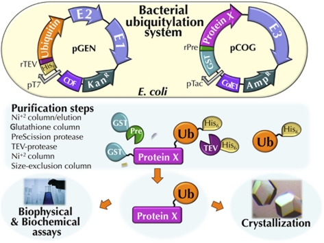
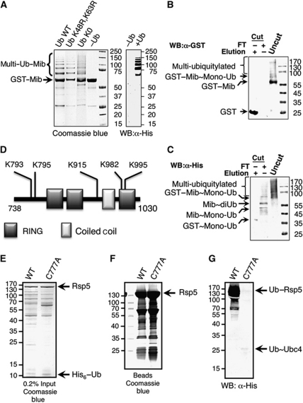
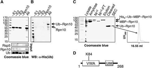
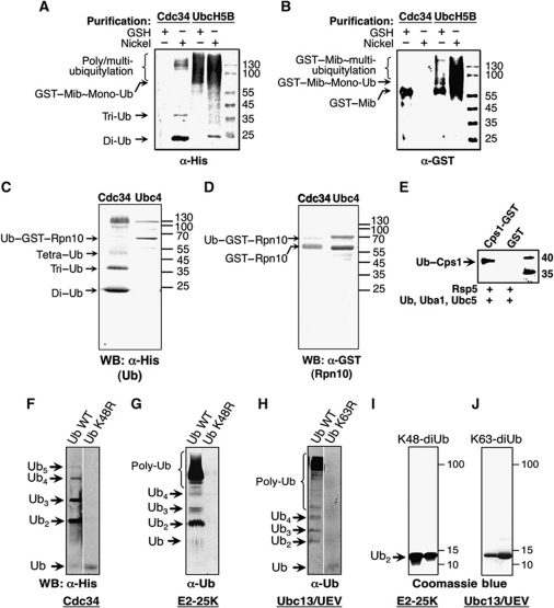
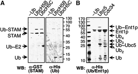
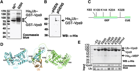
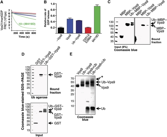
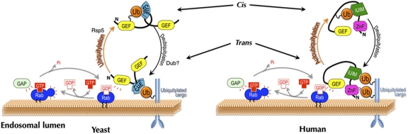
Covalent modification of proteins with ubiquitin (Ub) is widely implicated in the control of protein function and fate. Over 100 deubiquitylating enzymes rapidly reverse this modification, posing challenges to the biochemical and biophysical characterization of ubiquitylated proteins. We circumvented this limitation with a synthetic biology approach of reconstructing the entire eukaryotic Ub cascade in bacteria. Co-expression of affinity-tagged substrates and Ub with E1, E2 and E3 enzymes allows efficient purification of ubiquitylated proteins in milligram quantity. Contrary to in-vitro assays that lead to spurious modification of several lysine residues of Rpn10 (regulatory proteasomal non-ATPase subunit), the reconstituted system faithfully recapitulates its monoubiquitylation on lysine 84 that is observed in vivo. Mass spectrometry revealed the ubiquitylation sites on the Mind bomb E3 ligase and the Ub receptors Rpn10 and Vps9. Förster resonance energy transfer (FRET) analyses of ubiquitylated Vps9 purified from bacteria revealed that although ubiquitylation occurs on the Vps9-GEF domain, it does not affect the guanine nucleotide exchanging factor (GEF) activity in vitro. Finally, we demonstrated that ubiquitylated Vps9 assumes a closed structure, which blocks additional Ub binding. Characterization of several ubiquitylated proteins demonstrated the integrity, specificity and fidelity of the system, and revealed new biological findings.


Conflict of interest statement

The authors declare that they have no conflict of interest.

Figures

Figure 1
Figure 1
Bacterial system for expression and purification of ubiquitylated proteins. A scheme describing the ubiquitylation system and the steps for purifying ubiquitylated proteins from E. coli (see also Supplementary Table SIV).
Figure 2
Figure 2
Reconstituted E3 auto-ubiquitylation activity in bacteria. Auto-ubiquitylation of the RING and HECT E3-ligase protein families is shown. (AC) Bacterial lysates co-expressing of His6–Ub (or the indicated mutants), Uba1, UbcH5b (pGEN1), and GST–Mib (pCOG9) were purified on Ni+2 and/or GSH beads as indicated. (A) On the left, a Coomassie blue-stained SDS–PAGE of auto-ubiquitylated D. rerio (zebrafish) Mib, a representative RING-containing E3s ligase is shown. Wild-type Ub (lane 1 from the left), Ub K48R, K63R double mutant (lane 2), Ub K0 (lane 3) or without pGEN (lane 4). On the right, anti-His tag western blot of GSH purified Mib is shown, without or with pGEN1 (lanes 1 and 2, respectively). (B, C) Ubiquitylated Mib was purified on GSH (uncut), then GST tag was cleaved and the sample was bound to GSH beads. Flow through (FT) and elution (Elution) were analysed using western blot with anti-GST tag (B) or with anti-His tag (C). (D) Scheme of Mib ubiquitylation sites detected by mass spectrometry (see Supplementary Figure S1 and Supplementary Table SI). (EG) Auto-ubiquitylation of yeast Rsp5, a representative HECT-containing E3s. Bacterial lysates co-expressing of His6–Ub, Uba1, Ubc5 (pGEN5), and MBP–Rsp5 WT (pCOG3) or C777A mutant (pCOG7) were purified on amylose beads. (E) Input expression levels are shown. (F) Coomassie blue-stained SDS–PAGE of bound fraction is shown. (G) Western blot using anti-His tag of bound fraction is shown.
Figure 3
Figure 3
Purification of K84-monoubiquitylated Rpn10. (A, B) Rpn10 ubiquitylation in bacteria is shown. Bacterial lysates co-expressing of His6–Ub, Uba1, yeast Ubc5 (expressed from pGEN5) and GST–Rpn10 and MBP–Rsp5 (expressed from pCOG5) were purified on GSH beads. Systematic deletions of the ubiquitylation enzymes (−E1, −E2 and −E3) show that all are required for ubiquitylation. Only when all components are co-expressed, ubiquitylation is observed (All). (A) Coomassie blue-stained SDS–PAGE of GSH beads bound fraction is shown. Input expression levels of His6—Ub, MBP–Rsp5 and GST–Rpn10 are shown below. Typical expression levels of E1 and E2 are shown in Supplementary Figure S4. (B) Western blot analysis of same samples shown in (A) using anti-His tag antibody is shown. (C) Coomassie blue-stained SDS–PAGE of the purification steps of ubiquitylated Rpn10 is shown. Bacterial lysates co-expressing pGEN24, Rsp5 and MBP–Rpn10 expressed from pCOG30 and pCOG31, respectively, were purified on amylose-affinity matrix (amylose), followed by Ni+2 beads (Ni+2). The apo Rpn10 was removed by wash (FT-Ni+2), Rpn10 was eluted (elution); His6 and MBP tags were cleaved and removed (cleaved). The UV280 absorbance chromatogram output of the size exclusion column (SEC) is shown (right of the gel). (D) A scheme of Rpn10 ubiquitylation site detected by mass spectrometry (see Supplementary Figure S2 and Supplementary Table SII) is shown.
Figure 4
Figure 4
Specificity and fidelity of the ubiquitylation system in the bacteria. Specificity to cognate E2s by RING-containing (A and B) and by HECT-containing (C and D) E3 ligases is shown. (A, B) Bacterial lysates co-expressing GST–Mib (pCOG9) and His6–Ub, Uba1, and UbcH5b or Cdc34 (pGEN1 or pGEN8, respectively), were purified on Ni+2 and/or GSH beads as indicated. The purified proteins were resolved on SDS–PAGE and subjected to western blot analysis with anti-His tag or anti-GST antibodies as indicated. (A) Shows that Mib is ubiquitylated by UbcH5b as indicated in both purifications (Ni2+ and GSH). Free polyUb chains are shown in the Cdc34 lane that was purified on Ni2+, but not seen in the GSH purification. (B) The ubiquitylation of Mib by UbcH5b; Cdc34 does not ubiquitylates Mib (only apo Mib was detected) in the GSH purification is shown. (C, D) Bacterial lysates co-expressing MBP–Rsp5 and GST–Rpn10 (pCOG5) and His6–Ub, Uba1, and Ubc4 or Cdc34 (pGEN4 or pGEN8, respectively), were purified on Ni+2 beads. The purified proteins were resolved on SDS–PAGE and subjected to western blot with anti-His tag or anti-GST antibodies as indicated. (C) Shows that Rpn10 is ubiquitylated by Rsp5 in the presence of Ubc4. Free polyUb chains are shown in the Cdc34 lane blotted with anti-His. (D) Rsp5-dependent ubiquitylation of Rpn10 is clearly evident in the Ubc4 lane. In the Cdc34 lane, a faint band of ubiquitylated Rpn10 is seen. (E) E3:substrate specificity is shown. Rsp5-dependent ubiquitylation of Cps1 (N-terminal residues PVEKAPR) fused to GST (lane 1) versus GST alone (lane 2). (FH) Fidelity of the polyUb linkage is shown in free polyUb chains. Bacterial lysates co-expressing Ub, Uba1 and the specified E2s were subjected to western blot with the indicated antibodies. (F, G) Formation of K48-polyUb by Cdc34 or E2-25K, respectively and (H) the formation of K63-polyUb by UBC13/UEV1a are shown. (I, J) Coomassie blue-stained SDS–PAGE of fractions derived from size-exclusion chromatography of purified K48 and K63 diUb produced in the bacterial system is shown; proteins were purified using the Pickart's protocol (Pickart and Raasi, 2005) followed by gel filtration.
Figure 5
Figure 5
Yeast Epsin-1 (Ent1p) undergoes E3-independent ubiquitylation. (A) The bacterial system maintains recently reported (Hoeller et al, 2007) E3-independent ubiquitylation of mouse STAM2 with the UbcH5b or UbcH5c E2s. Bacterial lysates co-expressing of His6–Ub, Uba1, UbcH5b or UbcH5c (pGEN1 or pGEN3, respectively), and GST-STAM2 (pCOG11) were purified on GSH beads and subjected to western blot analysis with anti-GST (left) and anti-His tag (right) antibodies. (B) Western blot analysis showing that the yeast Epsin-1 (Ent1p) undergoes E3-independent ubiquitylation. Bacterial lysates co-expressing of His6–Ub, Uba1, yeast Ubc5 or yeast Cdc34 (pGEN6 or pGEN8, respectively), and His6–MBP–Ent1p (pCOG21) were purified on Ni+2 beads. Bound proteins were subjected to western blot analysis with anti-His tag antibody. Without pGEN, no ubiquitylation is observed (−Ub). With Ubc5, Ent1p ubiquitylation was observed (Ubc5). With Cdc34, no Ent1p ubiquitylation was detected (Cdc34). Nevertheless, formation of polyUb chains by Cdc34 was clearly observed, indicating it is active.
Figure 6
Figure 6
Identification of the ubiquitylation sites on yeast Vps9. Bacterial lysates co-expressing His6–Ub, Uba1, Ubc5 (pGEN5), Rsp5 and GST–Vps9 (pCOG2) were purified as described in Figure 1. (A) A Coomassie blue-stained SDS–PAGE of two sequential affinity chromatography purification steps (Ni+2 and GSH) of ubiquitylated Vps9 is shown. Since Vps9 is a dimer, the monoubiquitylated complex is composed of stoichiometry equal amounts of modified and non-modified proteins. (B) Western blot analysis of the purified protein with an anti-His-tag antibody is shown. (C) Identification of the ubiquitylation sites on Vps9 as found in mass spectrometry analysis (see Supplementary Table SIII and Supplementary Figure S3). (D) The ubiquitylation sites are projected on the structural model of Vps9. Vps9-GEF domain was modelled based on the structure of the human orthologue Rabex5 GEF domain (Delprato et al, 2004). The structure of the hinge region linking the GEF and the CUE domain was not modelled and is illustrated as dashed lines. The CUE:Ub complex (Prag et al, 2003) is shown where the C-terminus of Ub is conjugated to K328, one of the lysine residues that were found to be ubiquitylated. (E) Mutational analyses of specific lysine residues found to undergo ubiquitylation by mass spectrometry. Vps9 mutants were expressed as His6–MBP fusions (pCOG23–pCOG29) with Uba1, Ubc4 and Rsp5 (pGEN24 and pCOG30, respectively) and purified on Ni+2 beads. Western blot analysis with anti-His tag antibody of purified ubiquitylated Vps9 wild-type and mutant proteins is shown. Five single lysine to arginine mutants and a combined mutant harbours all five substitutions (5Xmut) are shown. All mutants underwent ubiquitylation with various levels compared with wild-type Vps9 (WT). Coomassie blue-stained SDS–PAGE with the indicated inputs (0.2%) is shown below.
Figure 7
Figure 7
Functionality of ubiquitylated Vps9. Purified ubiquitylated Vps9 derived from reconstituted ubiquitylation system in E. coli was used for GEF activity and Ub-binding assays. (A) Crude data of GEF activity analyses of Vps9, ubiquitylated Vps9, GEF:Rab interface double mutant (D251A/E288A) and CUE:Ub interface mutant (M419E) are shown (colour code as indicated in B). The GEF activity of ubiquitylated Vps9 was compared with that of wild type, D251A/E288A and M419D Vps9 in assays monitoring mantGDP release from Vps21 in the presence of excess GTP (1 mM). The concentrations of Vps9 and Vps21 were 100 nM and 2.5 μM, respectively. FRET between Vps21 and mantGDP (ex. 290 nm, em. 440 nm) was carried out to continuously assess Vps21•mantGDP binding over a 15-min incubation period (1 μM Vps9M419D-stimulated release is presented to indicate the range of Vps21•mantGDP release that could be achieved (× 10)). The rate of Vps9-stimulated mantGDP release relative to Vps21 alone is shown. (B) Averaged data of at least three independent experiments (as shown in A) are shown, and error bars indicate standard deviations. (CE) Direct evidences for close structure of ubiquitylated Vps9 are shown. Pull-down and crosslinking assays of Vps9 and ubiquitylated Vps9 with immobilized or free Ub are shown. (C) Ub-agarose beads pull-down assay with purified proteins expressed as MBP fusions (see Materials and methods). Only the apo-Vps9 binds Ub agarose while the Ub–MBP–Vps9 does not. (D) Ub agarose was used in the pull-down assay with E. coli cell extracts that expressed GST, GST–Vps9 or ubiquitylated GST–Vps9 (as indicated). Purified proteins from the same cell extracts (input) presented that all three are highly expressed. Only the apo-Vps9 binds Ub agarose. (E) Crosslinking assay of purified Ub, Vps9 and ubiquitylated Vps9 proteins is shown. A mild crosslinker (DSS) was used. Vps9 and ubiquitylated Vps9 do not show self-association (lanes 1 and 2 from the left). Vps9:Ub association represents formation of trans complex (lane 3). Ubiquitylated Vps9 does not bind free Ub (lane 4). No band of the predicted migration distance of Ub bound to ubiquitylated Vps9 is seen (as represented by *).
Figure 8
Figure 8
Model for the regulatory function of UBD-GEF proteins auto-ubiquitylation. A comparison is shown between models of yeast and of human GEF-containing Ub receptors of the endocytic pathway. The localization of human Rabex5 and of yeast Vps9 proteins is regulated by auto-ubiquitylation. Trans complexes with ubiquitylated transmembrane cargo are on the membrane of early endosomes. This trans interaction promotes the nucleotide exchange of the Rab5 protein family members that are anchored to the same membrane through a geranylgeranyl group. Auto-ubiquitylation of the GEF-containing Ub receptors blocks their UBDs in cis, and therefore sequesters them from the membrane. It is believed that a specific deubiquitylation enzyme (DUB) stimulates the cycle by reversing the ubiquitylation signal.

Similar articles

Cited by

References

    1. Azem A, Weiss C, Goloubinoff P (1998) Structural analysis of GroE chaperonin complexes using chemical cross-linking. Methods Enzymol 290: 253–268 - PubMed
    1. Baba D, Maita N, Jee JG, Uchimura Y, Saitoh H, Sugasawa K, Hanaoka F, Tochio H, Hiroaki H, Shirakawa M (2005) Crystal structure of thymine DNA glycosylase conjugated to SUMO-1. Nature 435: 979–982 - PubMed
    1. Bachmair A, Finley D, Varshavsky A (1986) In vivo half-life of a protein is a function of its amino-terminal residue. Science 234: 179–186 - PubMed
    1. Chatterjee C, McGinty RK, Fierz B, Muir TW (2010) Disulfide-directed histone ubiquitylation reveals plasticity in hDot1L activation. Nat Chem Biol 6: 267–269 - PubMed
    1. Chen J, Ai Y, Wang J, Haracska L, Zhuang Z (2010) Chemically ubiquitylated PCNA as a probe for eukaryotic translesion DNA synthesis. Nat Chem Biol 6: 270–272 - PubMed

Publication types

MeSH terms

Substances