. 2011 Dec 23;13(1):75-82.
doi: 10.1038/embor.2011.211.

Profilin1 is required for glial cell adhesion and radial migration of cerebellar granule neurons

Affiliations

Profilin1 is required for glial cell adhesion and radial migration of cerebellar granule neurons

Jan A Kullmann et al. EMBO Rep. .

Abstract

Cerebellar granule neurons (CGNs) exploit Bergmann glia (BG) fibres for radial migration, and cell-cell contacts have a pivotal role in this process. Nevertheless, little is known about the mechanisms that control CGN-BG interaction. Here we demonstrate that the actin-binding protein profilin1 is essential for CGN-glial cell adhesion and radial migration. Profilin1 ablation from mouse brains leads to a cerebellar hypoplasia, aberrant organization of cerebellar cortex layers and ectopic CGNs. Conversely, neuronal progenitor proliferation, tangential migration of neurons and BG morphology appear to be independent of profilin1. Our mouse data and the mapping of developmental neuropathies to the chromosomal region of PFN1 suggest a similar function for profilin1 in humans.


Conflict of interest statement

The authors declare that they have no conflict of interest.

Figures

Figure 1
Figure 1
Cerebellar hypoplasia in profilin1 mutants. (A) Immunoblot analysis proved efficient profilin1 deletion in mutant brains. Profilin2 levels were unchanged. Actin served as a loading control. (B) The cerebellum (arrow) was smaller in P60 mutants. Scale bar, 3 mm. (C,D) Nissl-stained parasagittal sections of a control and a mutant at P8 (C) and P60 (D). Asterisks depict folia 4/5 that were used for morphometric analysis (Table 1) and are shown at a higher magnification in Fig 2. Scale bar, 1 mm. (E) In adult huGE mice expressing GFP actin under the control of the profilin1 promoter, GFP signal was intense in the CGN-containing IGL and the ML, but not in the white matter. Scale bar, 50 μm. (F,G) Moreover, GFP signal was present in both calbindin-positive PC (F) and GFAP-positive BG (G). Scale bar, 20 μm in F and G. BG, Bergmann glia; CGN, cerebellar granule neuron; E, embryonic day; GFAP, glial fibrillary acidic protein; GFP, green fluorescent protein; IGL, internal granule cell layer; ML, molecular layer; P, postnatal day; PC, Purkinje cells.
Figure 2
Figure 2
Ectopic CGNs in profilin1 mutants. (A) Nissl staining revealed normal cerebellar cortex cytoarchitecture in P8 mutants. Scale bar, 50 μm in A–C. (B) In P12 mutants, layering of the cerebellar cortex was normal, whereas the ML cell density appeared higher. (C) Increased ML cell density, an irregularly shaped PCL (dashed lines) and gaps within the PCL (arrowhead) were noted in P60 mutants. (D) ML cell densities were significantly increased in mutants at P12 (controls: 4,702±222 cells per mm2; mutants: 7,411±206 cells per mm2; n=12 images from 6 mice; P<0.001) and P60 (controls: 1,975±150 cells per mm2; mutants: 3,397±119 cells per mm2; n=16/8; P<0.001), but not at P8 (controls: 5,182±221 cells per mm2; mutants: 6,069±387 cells per mm2; n=6/3; P=0.07). (E) NeuN immunoreactivity (green) and PI counterstaining (magenta) in the ML at P60. Scale bar, 40 μm. Yellow arrowheads indicate PI+/NeuN cells, whereas blue arrowheads indicate PI+/NeuN+ cells. (F) Although the number of PI+/NeuN cells was unchanged (controls: 1,971±88 cells per mm2; mutants: 1,789±115 cells per mm2; n=6/3; P=0.242), the number of PI+/NeuN+ cells was increased in the mutant ML (controls: 292±42 cells per mm2; mutants: 1,459±76 cells per mm2; P<0.001). CGN, cerebellar granule neuron; EGL, external granule cell layer; IGL, internal granule cell layer; ML, molecular layer; NeuN, neuron-specific nuclear protein; NS, not significant; P, postnatal day; PCL, Purkinje cell layer; PI, propidium iodide.
Figure 3
Figure 3
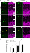
Impaired radial migration of cerebellar granule neurons (CGNs) in profilin1 mutants. (A–C) Representative images of BrdU+ cells (green) in the cerebellar cortex (A) 48 h, (B) 72 h and (C) 96 h after BrdU injection at postnatal day (P) 8. Sections were counterstained with PI (magenta). Examples for rounded (white arrowheads) and elongated (yellow arrowheads) CGNs located in the ML are shown in C. Scale bar, 50 μm for AC. (D) Mean migration distance of BrdU+ cells was reduced in mutants at all time points (48 h: controls: 79.4±3.4 μm; mutants:55.0±4.4 μm; n=6 images from 3 mice; P<0.01; 72 h: controls: 109.7±3.7 μm; mutants: 97.6±8.6 μm; n=6/3; P<0.05; 96 h: controls: 169.5±8.7 μm; mutants: 111.5±7.5 μm; n=6/3; P<0.001). BrdU, 5-bromo-2-deoxyuridine; CGN, cerebellar granule neuron; EGL, external granule cell layer; IGL, internal granule cell layer; ML, molecular layer; PI, propidium iodide.
Figure 4
Figure 4
Profilin1 activity in CGNs and GC is required for CGN–GC adhesion and radial migration. (A) Representative time-lapse images showing reduced migration of profilin1-deficient CGNs (CGN−/−) along processes of control GC (GC+/+) and of control CGN (CGN+/+) along processes of profilin1-deficient GC (GC−/−). Scale bar, 10 μm. (B) When profilin1 was absent in CGNs (CGN−/−/GC+/+) or in GC (CGN+/+/GC−/−), mean CGN migration velocity was significantly reduced (CGN+/+/GC+/+: 56.3±4.5 μm/h, n=17; CGN−/−/GC+/+: 38.4±2.7 μm h−1, n=21, P<0.01; CGN+/+/GC−/−: 39.7±3.1 μm h−1, n=16, P<0.01). (C) Compared with adhesion of control CGNs and control GC (CGN+/+/GC+/+: 22.6±1.7 cells per mm2; n=20), CGN–GC adhesion was significantly impaired when profilin1 was absent from either CGNs (CGN−/−/GC+/+: 11.6±1.0 cells per mm2; n=11; P<0.001) or GC (CGN+/+/GC−/−: 7.4±1.5 cells per mm2; n=8; P<0.001). (D) Mena and vinculin immunoreactivity in isolated CGN+/+ and CGN−/− attached to processes of GC+/+ (GCP). Scale bar, 5 μm. Dashed line in the upper left micrograph exemplarily illustrates CGN–GC junctions as chosen for further analyses. (E) Mean Mena and vinculin fluorescence intensities in CGN–GC junctions were reduced when profilin1 was absent from CGNs (Mena: CGN+/+/GC+/+: 48.0±5.1 AU; CGN−/−/GC+/+: 26.3±4.5 AU; n=8; P<0.01; vinculin: CGN+/+/GC+/+: 37.1±3.8 AU; CGN−/−/GC+/+: 19.5±3.7 AU; n=9; P<0.01). AU, arbitrary units; CGN, cerebellar granule neuron; GC, glial cell.

References

    1. Adams NC, Tomoda T, Cooper M, Dietz G, Hatten ME (2002) Mice that lack astrotactin have slowed neuronal migration. Development 129: 965–972 - PubMed
    1. Arregui CO, Carbonetto S, McKerracher L (1994) Characterization of neural cell adhesion sites: point contacts are the sites of interaction between integrins and the cytoskeleton in PC12 cells. J Neurosci 14: 6967–6977 - PMC - PubMed
    1. Ayala R, Shu T, Tsai LH (2007) Trekking across the brain: the journey of neuronal migration. Cell 128: 29–43 - PubMed
    1. Bellenchi GC, Gurniak CB, Perlas E, Middei S, Ammassari-Teule M, Witke W (2007) N-cofilin is associated with neuronal migration disorders and cell cycle control in the cerebral cortex. Genes Dev 21: 2347–2357 - PMC - PubMed
    1. Bershadsky A (2004) Magic touch: how does cell–cell adhesion trigger actin assembly? Trends Cell Biol 14: 589–593 - PubMed

Publication types

LinkOut - more resources