

doi: 10.1038/embor.2011.209.
CD8αα and -αβ isotypes are equally recruited to the immunological synapse through their ability to bind to MHC class I
Affiliations
- PMID: 22081144
- PMCID: PMC3245696
- DOI: 10.1038/embor.2011.209
Item in Clipboard
CD8αα and -αβ isotypes are equally recruited to the immunological synapse through their ability to bind to MHC class I
EMBO Rep.
.
Abstract
Bimolecular fluorescence complementation was used to engineer CD8 molecules so that CD8αα and CD8αβ dimers can be independently visualized on the surface of a T cell during antigen recognition. Using this approach, we show that CD8αα is recruited to the immunological synapse almost as well as CD8αβ, but because the kinase Lck associates preferentially with CD8αβ in lipid rafts, CD8αα is the weaker co-receptor. During recognition of the strong CD8αα ligand H2-TL, CD8αα is preferentially recruited. Thus, recruitment of the two CD8 species correlates with their relative binding to the available ligands, rather than with the co-receptor functions of the CD8 species.
Conflict of interest statement
The authors declare that they have no conflict of interest.
Figures
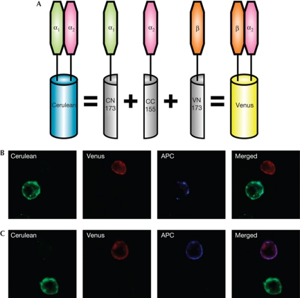
Principle of the bimolecular fluorescence complementation (BiFC) assay and specific CD8α1 and CD8β detection. (A) CD8α2–CC155 forms CD8αα when expressed with CD8α1–CN173, or CD8αβ when expressed with CD8β–CN173. Note that non-fluorescent CD8αα or CD8αβ can also be formed from CD8α1 or CD8α2 homodimers, or from CD8α1–CD8β heterodimers. (B,C) Cells expressing only the fluorescent molecules CD8α2α1 (Cerulean: shown as green) or CD8α2β (Venus, shown as red) were mixed at a 1:1 ratio and stained with allophycocyanin (APC)–anti-CD8α1 (B) or APC–anti-CD8β (C). The antibody staining is shown in blue.
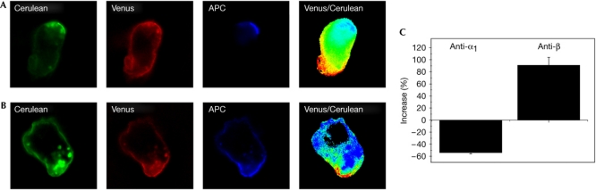
Separate capping of CD8αα and CD8αβ. (A,B) Representative images of co-capping with anti-CD8α1 (anti-Ly2.1) (A) or anti-CD8β (B) monoclonal antibodies on cells expressing all three bimolecular fluorescence complementation (BiFC) constructs. On panels labelled Venus/Cerulean, the intensity ratio of Venus/Cerulean is represented using a high/low scale in which blue indicates a lower ratio value (more CD8α2α1) and red a higher ratio value (more CD8α2β). (C) Shows quantification of the differential capping of CD8α2α1 and CD8α2β as percentage of increase. Negative values reflect preferential recruitment of CD8α2α1 and positive values the preferential recruitment of CD8α2β. Error bars show s.e.m., n=16 for anti-α1 and n=30 for anti-β. APC, allophycocyanin.
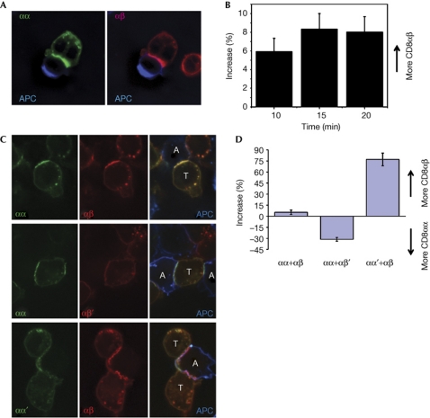
CD8αα or αβ recruitment to the IS depends on the ability to bind to MHC-I. (A) T cell expressing CD8αα-Cerulean and CD8αβ-Venus interacting with a Cy5-labelled antigen-presenting cell (APC). For clarity, Cerulean and Venus channels merged with the Cy5 channel are presented separately. (B) Time course of differential recruitment of CD8αα-Cerulean and CD8αβ-Venus in cells responding to RMA-S cells presenting Kb-OVA. Graph shows the percentage of increase ±s.e.m., n⩾20. (C) T cells expressing CD8αα+CD8αβ, CD8αα+CD8αβ′ or CD8αα′+CD8αβ fluorescent proteins during interaction with ovalbumin (OVA) peptide-loaded EL4 cells. Cerulean and Venus channels are presented separately (left and centre) and merged with Cy5 (APC) channel in right panels. T cells and APCs are labelled on the merged image (T and A, respectively). (D) Preferential recruitment of wild-type co-receptor species compared with non-major histocompatibility complex-I-binding mutants to the immunological synapse (IS). Negative values reflect preferential recruitment of CD8αα and positive values the preferential recruitment of CD8αβ. Graph shows the percentage of increase ±s.e.m. n=17, 18 and 15.
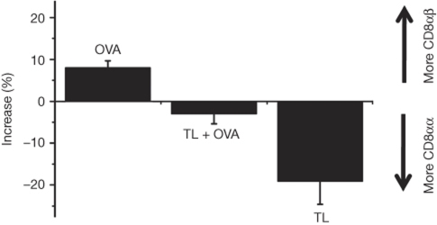
Ligand dependence of the differential recruitment of CD8αα and CD8αβ. Data are presented for T cells expressing CD8αα-Cerulean and CD8αβ-Venus responding to RMA-S cells presenting Kb-OVA (OVA), RMA-S cells expressing thymus leukaemia antigen (H2-TL or TL) and also presenting Kb-OVA (TL+OVA) and RMA-S cells expressing TL alone (TL). Error bars represent s.d. n=25 for each group.
Similar articles
-
The murine CD94/NKG2 ligand, Qa-1b, is a high-affinity, functional ligand for the CD8αα homodimer.J Biol Chem. 2020 Mar 6;295(10):3239-3246. doi: 10.1074/jbc.RA119.010509. Epub 2020 Jan 28. J Biol Chem. 2020. PMID: 31992596 Free PMC article.
-
The CD8 isoform CD8alphaalpha is not a functional homologue of the TCR co-receptor CD8alphabeta.Curr Opin Immunol. 2004 Jun;16(3):264-70. doi: 10.1016/j.coi.2004.03.015. Curr Opin Immunol. 2004. PMID: 15134773 Review.
-
Late postnatal expansion of self-reactive CD8alphaalpha+ intestinal intraepithelial lymphocytes in mice.Autoimmunity. 2004 Dec;37(8):537-47. doi: 10.1080/08916930400027094. Autoimmunity. 2004. PMID: 15763916
-
MHC class I allele dosage alters CD8 expression by intestinal intraepithelial lymphocytes.J Immunol. 2001 Sep 1;167(5):2561-8. doi: 10.4049/jimmunol.167.5.2561. J Immunol. 2001. PMID: 11509596
-
Doubting the TCR coreceptor function of CD8alphaalpha.Immunity. 2008 Feb;28(2):149-59. doi: 10.1016/j.immuni.2008.01.005. Immunity. 2008. PMID: 18275828 Review.
Cited by
-
CD8αα+T cells exert a pro-inflammatory role in patients with psoriasis.Skin Health Dis. 2021 Nov 16;1(4):e64. doi: 10.1002/ski2.64. eCollection 2021 Dec. Skin Health Dis. 2021. PMID: 35663772 Free PMC article.
-
A ternary complex comprising transportin1, Rab8 and the ciliary targeting signal directs proteins to ciliary membranes.J Cell Sci. 2016 Oct 15;129(20):3922-3934. doi: 10.1242/jcs.194019. Epub 2016 Sep 15. J Cell Sci. 2016. PMID: 27633000 Free PMC article.
-
The kinase occupancy of T cell coreceptors reconsidered.Proc Natl Acad Sci U S A. 2022 Dec 6;119(49):e2213538119. doi: 10.1073/pnas.2213538119. Epub 2022 Dec 1. Proc Natl Acad Sci U S A. 2022. PMID: 36454761 Free PMC article.
-
The molecular determinants of CD8 co-receptor function.Immunology. 2012 Oct;137(2):139-48. doi: 10.1111/j.1365-2567.2012.03625.x. Immunology. 2012. PMID: 22804746 Free PMC article. Review.
-
Initiation of TCR phosphorylation and signal transduction.Front Immunol. 2011 Dec 7;2:72. doi: 10.3389/fimmu.2011.00072. eCollection 2011. Front Immunol. 2011. PMID: 22566861 Free PMC article.
References
-
- Arcaro A, Gregoire C, Boucheron N, Stotz S, Palmer E, Malissen B, Luescher IF (2000) Essential role of CD8 palmitoylation in CD8 coreceptor function. J Immunol 165: 2068–2076 - PubMed
-
- Attinger A, Devine L, Wang-Zhu Y, Martin D, Wang JH, Reinherz EL, Kronenberg M, Cheroutre H, Kavathas P (2005) Molecular basis for the high affinity interaction between the thymic leukemia antigen and the CD8αα molecule. J Immunol 174: 3501–3507 - PubMed
Publication types
MeSH terms
Substances
Grants and funding
LinkOut - more resources
Full Text Sources
Molecular Biology Databases
Research Materials
Miscellaneous
