. 2012 Jun;227(2):165-74.
doi: 10.1002/path.3960. Epub 2012 Feb 17.

Chronic inhibition of endoplasmic reticulum stress and inflammation prevents ischaemia-induced vascular pathology in type II diabetic mice

Affiliations

Chronic inhibition of endoplasmic reticulum stress and inflammation prevents ischaemia-induced vascular pathology in type II diabetic mice

Ali Amin et al. J Pathol. 2012 Jun.

Abstract

Endoplasmic reticulum (ER) stress and inflammation are important mechanisms that underlie many of the serious consequences of type II diabetes. However, the role of ER stress and inflammation in impaired ischaemia-induced neovascularization in type II diabetes is unknown. We studied ischaemia-induced neovascularization in the hind-limb of 4-week-old db - /db- mice and their controls treated with or without the ER stress inhibitor (tauroursodeoxycholic acid, TUDCA, 150 mg/kg per day) and interleukin-1 receptor antagonist (anakinra, 0.5 µg/mouse per day) for 4 weeks. Blood pressure was similar in all groups of mice. Blood glucose, insulin levels, and body weight were reduced in db - /db- mice treated with TUDCA. Increased cholesterol and reduced adiponectin in db - /db- mice were restored by TUDCA and anakinra treatment. ER stress and inflammation in the ischaemic hind-limb in db - /db- mice were attenuated by TUDCA and anakinra treatment. Ischaemia-induced neovascularization and blood flow recovery were significantly reduced in db - /db- mice compared to control. Interestingly, neovascularization and blood flow recovery were restored in db - /db- mice treated with TUDCA or anakinra compared to non-treated db - /db- mice. TUDCA and anakinra enhanced eNOS-cGMP, VEGFR2, and reduced ERK1/2 MAP-kinase signalling, while endothelial progenitor cell number was similar in all groups of mice. Our findings demonstrate that the inhibition of ER stress and inflammation prevents impaired ischaemia-induced neovascularization in type II diabetic mice. Thus, ER stress and inflammation could be potential targets for a novel therapeutic approach to prevent impaired ischaemia-induced vascular pathology in type II diabetes.


Conflict of interest statement

No conflicts of interest were declared.

Figures

Figure 1
Figure 1
(A) Systolic arterial blood pressure in all groups measured by the tail cuff methods. n = 7. (B) Body weight in control (CTR) and db−/db− mice treated with and without TUDCA (TUD) and anakinra (Ana). n = 7. *p < 0.05 for CTR versus db−/db−; #p < 0.05 for db−/db− versus db−/db− treated with TUDCA. (C) Blood glucose levels in all groups. n = 7. *p < 0.05 for CTR and db−/db− mice treated with TUDCA versus db−/db− with and without anakinra. (D) Insulin level in all groups. n = 7. *p < 0.05 for CTR and db−/db− treated with TUDCA versus db−/db− with and without anakinra. (E) Cholesterol levels in all groups. n = 4. *p < 0.05 for db−/db− versus CTR, db−/db− + TUDCA and Ana. (F) Adiponectin levels in all groups. n = 4. *p < 0.05 for db−/db− versus CTR, db−/db− + TUDCA and Ana.
Figure 2
Figure 2
(A) ATF4 and CHOP mRNA levels determined using real-time RT-PCR in ischaemic hind-limb from all groups. n = 5. *p < 0.05 for db−/db− versus control (CTR) mice and db−/db− mice with and without TUDCA and anakinra (Ana). (B) Immunostaining of macrophages in the ischaemic hind-limb from all groups. (This figure is representative of n = 5.) (C) CRP levels in all groups. n = 4. *p < 0.05 for db−/db− versus CTR, db−/db− + TUDCA and Ana.
Figure 3
Figure 3
(A) Blood flow recovery measured with a Moor LPDI laser in the ischaemic hind-limb of control (CTR) and db−/db− mice with or without TUDCA and anakinra (Ana) before (Pre) and after surgery, and once a week for 4 weeks. This picture is representative of n = 8. (B) Quantitative data for blood flow recovery in all groups of mice. n = 8. *p < 0.05 for db−/db− mice versus control (CTR) and db−/db− mice + TUDCA and anakinra (Ana). (C) Blood flow recovery measured in ischaemic hind-limb of control (CTR) mice and CTR mice locally injected with tunicamycin (TUN) for 4 weeks or interleukin-1 (IL-1) for 2 weeks. n = 4. *p < 0.05 for CTR versus CTR + TUN or IL-1.
Figure 4
Figure 4
(A) Microangiography performed at the end of the treatment in all groups of mice [control (CTR), and db−/db− with and without TUDCA and anakinra (Ana)]. Contrast medium (barium sulphate, 0.5 μg/ml) was injected into the abdominal aorta and angiography of the right and left hind-limbs was assessed with digital X-ray-acquired images. Images were then assembled to obtain a composite view of the hind-limb. The image is representative of n = 6. (B) Quantitative data (score in %) showing ischaemic hind-limb vessel density using Multi Gauge (Fujifilm) by selecting the same area for measurement in all groups. n = 6. *p < 0.05 for db−/db− mice versus control (CTR) and db−/db− mice + TUDCA and anakinra (Ana).
Figure 5
Figure 5
Example of immunostaining with specific antibodies for α-actin (red staining, white arrows) and CD31 (green staining; capillaries). Yellow staining (yellow arrows) represents arterioles which ‘merge between green and red’. Quantitative data of CD31 expression in all groups. n = 5. *p < 0.05 for db−/db− mice versus control (CTR) and db−/db− mice + TUDCA and anakinra (Ana).
Figure 6
Figure 6
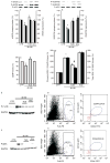
Western blot analysis and quantitative data showing total (T) and phosphorylated (P) eNOS in the ischaemic hind-limb from all groups (A); total (T) and phosphorylated (P) VEGFR2 in ischaemic hind-limb from all groups (B); and total (T) and phosphorylated (P) ERK1/2 MAP-kinase in ischaemic hind-limb from all groups (D). n = 6. *p < 0.05 for db−/db− mice versus control (CTR) and db−/db− mice + TUDCA and anakinra (Ana). (C) cGMP levels in ischaemic hind-limb from all groups. n = 6. *p < 0.05 for db−/db− mice versus control (CTR) and db−/db− mice + TUDCA and anakinra (Ana). (E, F) Phosphorylated C-JNK (P-C-JNK) and MEK (P-MEK) in ischaemic hind-limb in all groups of mice. (G, H) Flow cytometry for EPCs in bone marrow from db−/db− and control mice.

References

    1. Harding HP, Ron D. Endoplasmic reticulum stress and the development of diabetes: a review. Diabetes. 2002;51(Suppl 3):S455–S461. - PubMed
    1. Mannava K, Money SR. Current management of peripheral arterial occlusive disease: a review of pharmacologic agents and other interventions. Am J Cardiovasc Drugs. 2007;7:59–66. - PubMed
    1. Cooke JP. NO and angiogenesis. Atheroscler Suppl. 2003;4:53–60. - PubMed
    1. Bernardini G, Ribatti D, Spinetti G, et al. Analysis of the role of chemokines in angiogenesis. J Immunol Methods. 2003;273:83–101. - PubMed
    1. Morbidelli L, Donnini S, Ziche M. Role of nitric oxide in the modulation of angiogenesis. Curr Pharm Des. 2003;9:521–530. - PubMed

Publication types

MeSH terms