

Galactose differentially modulates lunatic and manic fringe effects on Delta1-induced NOTCH signaling
- PMID: 22081605
- PMCID: PMC3249100
- DOI: 10.1074/jbc.M111.317578
Galactose differentially modulates lunatic and manic fringe effects on Delta1-induced NOTCH signaling
Abstract
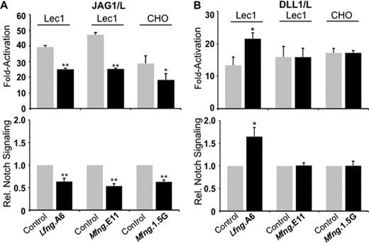
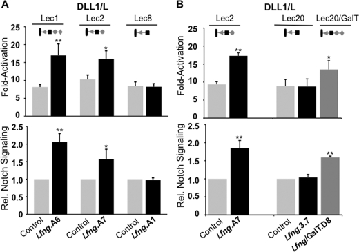
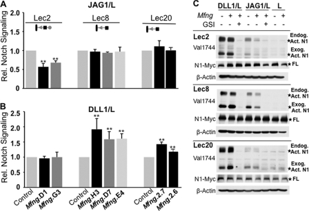
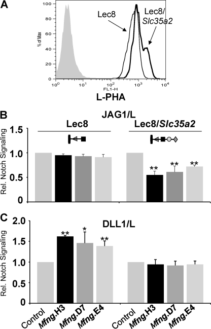
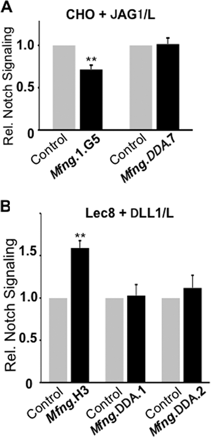
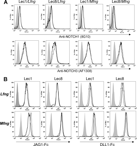
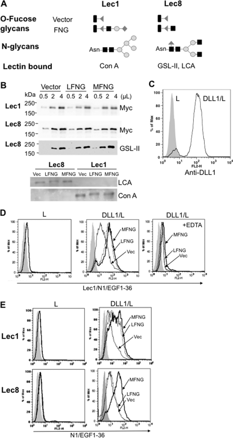
NOTCH signaling induced by Delta1 (DLL1) and Jagged1 (JAG1) NOTCH ligands is modulated by the β3N-acetylglucosaminyl transferase Fringe. LFNG (Lunatic Fringe) and MFNG (Manic Fringe) transfer N-acetylglucosamine (GlcNAc) to O-fucose attached to EGF-like repeats of NOTCH receptors. In co-culture NOTCH signaling assays, LFNG generally enhances DLL1-induced, but inhibits JAG1-induced, NOTCH signaling. In mutant Chinese hamster ovary (CHO) cells that do not add galactose (Gal) to the GlcNAc transferred by Fringe, JAG1-induced NOTCH signaling is not inhibited by LFNG or MFNG. In mouse embryos lacking B4galt1, NOTCH signaling is subtly reduced during somitogenesis. Here we show that DLL1-induced NOTCH signaling in CHO cells was enhanced by LFNG, but this did not occur in either Lec8 or Lec20 CHO mutants lacking Gal on O-fucose glycans. Lec20 mutants corrected with a B4galt1 cDNA became responsive to LFNG. By contrast, MFNG promoted DLL1-induced NOTCH signaling better in the absence of Gal than in its presence. This effect was reversed in Lec8 cells corrected by expression of a UDP-Gal transporter cDNA. The MFNG effect was abolished by a DDD to DDA mutation that inactivates MFNG GlcNAc transferase activity. The binding of soluble NOTCH ligands and NOTCH1/EGF1-36 generally reflected changes in NOTCH signaling caused by LFNG and MFNG. Therefore, the presence of Gal on O-fucose glycans differentially affects DLL1-induced NOTCH signaling modulated by LFNG versus MFNG. Gal enhances the effect of LFNG but inhibits the effect of MFNG on DLL1-induced NOTCH signaling, with functional consequences for regulating the strength of NOTCH signaling.
Figures
Similar articles
-
Fringe glycosyltransferases differentially modulate Notch1 proteolysis induced by Delta1 and Jagged1.Mol Biol Cell. 2005 Feb;16(2):927-42. doi: 10.1091/mbc.e04-07-0614. Epub 2004 Dec 1. Mol Biol Cell. 2005. PMID: 15574878 Free PMC article.
-
Disrupting Jagged1-Notch signaling impairs spatial memory formation in adult mice.Neurobiol Learn Mem. 2013 Jul;103:39-49. doi: 10.1016/j.nlm.2013.03.001. Epub 2013 Apr 6. Neurobiol Learn Mem. 2013. PMID: 23567106
-
Fringe differentially modulates Jagged1 and Delta1 signalling through Notch1 and Notch2.Nat Cell Biol. 2000 Aug;2(8):515-20. doi: 10.1038/35019553. Nat Cell Biol. 2000. PMID: 10934472
-
Regulation of Notch signaling during T- and B-cell development by O-fucose glycans.Immunol Rev. 2009 Jul;230(1):201-15. doi: 10.1111/j.1600-065X.2009.00791.x. Immunol Rev. 2009. PMID: 19594638 Review.
-
Regulation of myeloid and lymphoid cell development by O-glycans on Notch.Front Mol Biosci. 2022 Nov 4;9:979724. doi: 10.3389/fmolb.2022.979724. eCollection 2022. Front Mol Biosci. 2022. PMID: 36406268 Free PMC article. Review.
Cited by
-
A compendium of Androgen Receptor Variant 7 target genes and their role in Castration Resistant Prostate Cancer.Front Oncol. 2023 Mar 1;13:1129140. doi: 10.3389/fonc.2023.1129140. eCollection 2023. Front Oncol. 2023. PMID: 36937454 Free PMC article. Review.
-
Novel roles for O-linked glycans in protein folding.Glycoconj J. 2014 Oct;31(6-7):417-26. doi: 10.1007/s10719-014-9556-4. Glycoconj J. 2014. PMID: 25186198 Free PMC article.
-
Structural Divergence in O-GlcNAc Glycans Displayed on Epidermal Growth Factor-like Repeats of Mammalian Notch1.Molecules. 2018 Jul 17;23(7):1745. doi: 10.3390/molecules23071745. Molecules. 2018. PMID: 30018219 Free PMC article.
-
Deciphering the Fringe-Mediated Notch Code: Identification of Activating and Inhibiting Sites Allowing Discrimination between Ligands.Dev Cell. 2017 Jan 23;40(2):193-201. doi: 10.1016/j.devcel.2016.12.013. Epub 2017 Jan 12. Dev Cell. 2017. PMID: 28089369 Free PMC article.
-
Galectin-3 inhibits osteoblast differentiation through notch signaling.Neoplasia. 2014 Nov 20;16(11):939-49. doi: 10.1016/j.neo.2014.09.005. eCollection 2014 Nov. Neoplasia. 2014. PMID: 25425968 Free PMC article.
References
-
- Artavanis-Tsakonas S., Muskavitch M. A. (2010) Curr. Top. Dev. Biol. 92, 1–29 - PubMed
-
- Bolós V., Grego-Bessa J., de la Pompa J. L. (2007) Endocr. Rev. 28, 339–363 - PubMed
-
- Rampal R., Luther K. B., Haltiwanger R. S. (2007) Curr. Mol. Med. 7, 427–445 - PubMed
-
- Koch U., Radtke F. (2010) Curr. Top. Dev. Biol. 92, 411–455 - PubMed
Publication types
MeSH terms
Substances
Grants and funding
LinkOut - more resources
Full Text Sources
Research Materials
Miscellaneous
