. 2012 Jan 2;287(1):408-417.
doi: 10.1074/jbc.M111.261818. Epub 2011 Nov 11.

Fat-derived factor omentin stimulates endothelial cell function and ischemia-induced revascularization via endothelial nitric oxide synthase-dependent mechanism

Affiliations

Fat-derived factor omentin stimulates endothelial cell function and ischemia-induced revascularization via endothelial nitric oxide synthase-dependent mechanism

Sonomi Maruyama et al. J Biol Chem. .

Abstract

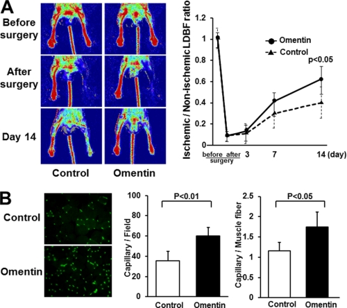
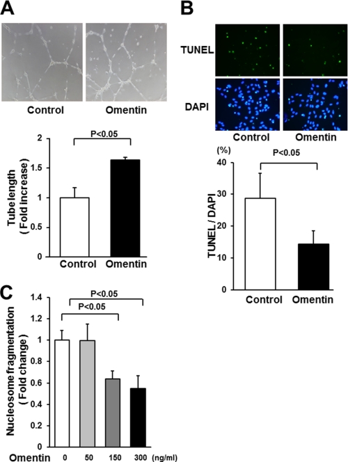
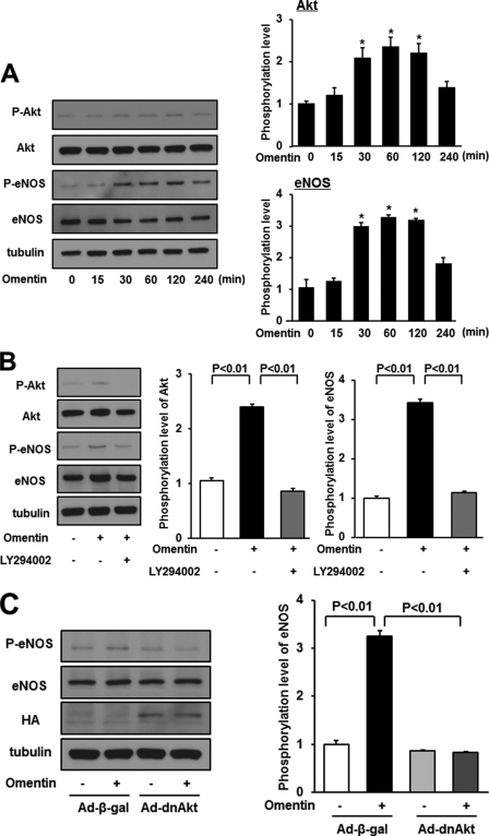
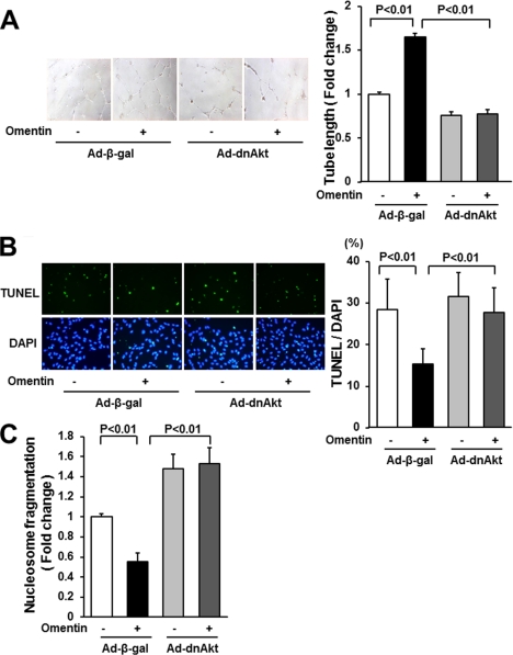
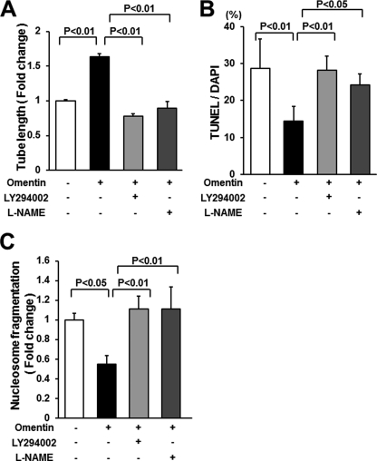
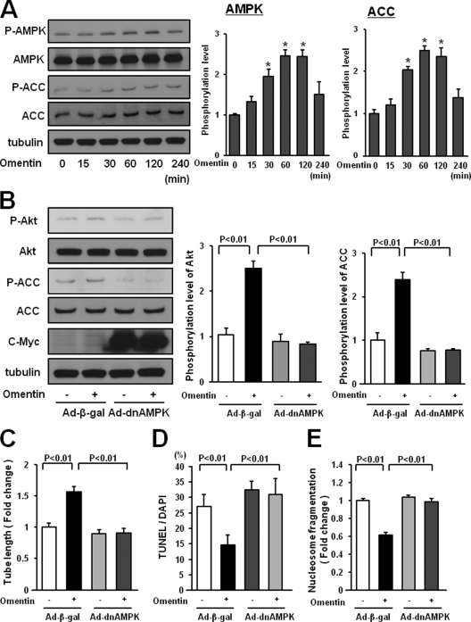
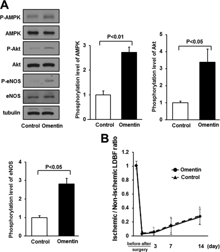
Obesity-related diseases are associated with vascular dysfunction and impaired revascularization. Omentin is a fat-derived secreted protein, which is down-regulated in association with obese complications. Here, we investigated whether omentin modulates endothelial cell function and revascularization processes in vitro and in vivo. Systemic delivery of an adenoviral vector expressing omentin (Ad-omentin) enhanced blood flow recovery and capillary density in ischemic limbs of wild-type mice in vivo, which were accompanied by increased phosphorylation of Akt and endothelial nitric oxide synthase (eNOS). In cultured human umbilical vein endothelial cells (HUVECs), a physiological concentration of recombinant omentin protein increased differentiation into vascular-like structures and decreased apoptotic activity under conditions of serum starvation. Treatment with omentin protein stimulated the phosphorylation of Akt and eNOS in HUVECs. Inhibition of Akt signaling by treatment with dominant-negative Akt or LY294002 blocked the stimulatory effects of omentin on differentiation and survival of HUVECs and reversed omentin-stimulated eNOS phosphorylation. Pretreatment with the NOS inhibitor also reduced the omentin-induced increase in HUVEC differentiation and survival. Omentin protein also stimulated the phosphorylation of AMP-activated protein kinase in HUVECs. Transduction with dominant-negative AMP-activated protein kinase diminished omentin-induced phosphorylation of Akt and omentin-stimulated increase in HUVEC differentiation and survival. Of importance, in contrast to wild-type mice, systemic administration of Ad-omentin did not affect blood flow in ischemic muscle in eNOS-deficient mice in vivo. These data indicate that omentin promotes endothelial cell function and revascularization in response to ischemia through its ability to stimulate an Akt-eNOS signaling pathway.


Figures

FIGURE 1.
FIGURE 1.
Omentin promotes perfusion recovery and capillary vessel formation of ischemic limbs in mice in vivo. Ad-omentin or Ad-β-gal (control) was injected into the jugular vein of WT mice (2 × 10⁷ pfu in each group) 3 days prior to surgery. A, representative LDBF images in ischemic limb of Ad-omentin-treated or control mice (left panels). Quantitative analysis of the ischemic/non-ischemic LDBF ratio in Ad-omentin-treated and control WT mice before surgery, after surgery, and on postoperative days 3, 7, and 14 (right panel). Results are shown as the mean ± S.D. (n = 7). B, fluorescence staining of ischemic tissues with anti-CD31 monoclonal antibody (green; left panel). Quantitative analysis of capillary density in Ad-omentin-treated and control mice on postoperative day 14. Capillary density was expressed as the number of capillaries per high power field (×400, middle panel) and capillaries per muscle fiber (right panel). Results are shown as the mean ± S.D. (n = 5).
FIGURE 2.
FIGURE 2.
Omentin promotes endothelial cell differentiation and survival in vitro. A, endothelial cell network formation following stimulation with omentin. After 18 h of serum deprivation, HUVECs were treated with recombinant omentin protein (300 ng/ml) or vehicle followed by culture on Matrigel-coated dishes. Representative photomicrographs of HUVEC differentiation into network structures are shown (upper panel). Quantitative analyses of network tube length are shown (bottom panel) (n = 3). B, omentin reduces the frequency of TUNEL-positive HUVECs. HUVECs were treated with omentin protein (300 ng/ml) or vehicle followed by 48 h of incubation with serum-free media. Apoptotic nuclei were identified by TUNEL staining (green), and total nuclei were identified by 4′,6-diamidino-2-phenylindole counterstaining (blue). Representative photomicrographs of TUNEL-positive HUVECs are shown (upper panels). Quantitative analyses of the frequency of TUNEL-positive HUVECs are shown (bottom panel) (n = 5). C, omentin inhibits the degree of nucleosome fragmentation of HUVECs. Nucleosome fragmentation was assessed by enzyme-linked immunosorbent assay. Results are expressed relative to the values compared with control. Results are shown as the mean ± S.D. (n = 5).
FIGURE 3.
FIGURE 3.
Omentin stimulates the phosphorylation of eNOS in endothelial cells via activation of Akt. A, time-dependent changes in the phosphorylation of Akt and eNOS signaling in endothelial cells following stimulation with omentitn (left). Changes in the phosphorylation of Akt (P-Akt) and eNOS (P-eNOS) following omentin treatment were determined by Western blot analysis. Representative blots are shown. Relative phosphorylation levels of Akt and eNOS were quantified by using ImageJ software. Immunoblots were normalized to an α-tubulin signal. Results are shown as the mean ± S.D. (n = 3). B, effect of LY294002 on omentin-stimulated eNOS phosphorylation. HUVECs were pretreated with LY294002 (50 μmol/L) or vehicle (dimethyl sulfoxide) for 60 min and treated with omentin (300 ng/ml) or vehicle for 60 min. P-Akt and P-eNOS were determined by Western blot analysis. Relative phosphorylation levels of Akt and eNOS were quantified by using ImageJ software. Immunoblots were normalized to α-tubulin signal. Results are shown as the mean ± S.D. (n = 3). C, role of Akt in regulation of omentin-induced eNOS signaling. HUVECs were transduced with Ad-dnAkt or Ad-β-gal 24 h before serum starvation. After 16 h serum starvation, cells were treated with omentin (300 ng/ml) for 60 min. P-eNOS was determined by Western blot analysis.
FIGURE 4.
FIGURE 4.
Akt signaling participates in endothelial cell responses to omentin. A, Akt signaling is essential for omentin-mediated endothelial cell differentiation. HUVECs were transduced with Ad-dnAkt or Ad-β-gal. After 18 h of serum starvation, a Matrigel assay was performed. Cells were treated with omentin (300 ng/ml) or vehicle. Representative photomicrographs of vascular-like tube formation are shown (left panels). Quantitative analyses of the network tube length are shown (right panel). Results are shown as the mean ± S.D. (n = 5). B and C, involvement of Akt in omentin-induced endothelial cell survival. After transduction with Ad-dnAkt or Ad-β-gal, cells were incubated in serum-free media for 48 h. Cells were treated with omentin (300 ng/ml) or vehicle. Apoptotic nuclei were identified by TUNEL staining (green), and total nuclei were identified by 4′,6-diamidino-2-phenylindole counterstaining (blue; B). Representative photomicrographs of TUNEL-positive HUVECs are shown (B, left panels). Quantitative analyses of the frequency of TUNEL-positive HUVECs are shown (B, right panel). Nucleosome fragmentation was assessed by enzyme-linked immunosorbent assay (C). Results are shown as the mean ± S.D. (n = 5).
FIGURE 5.
FIGURE 5.
PI3K and eNOS contribute to omentin-induced endothelial cell responses. A, contribution of PI3K and eNOS to omentin-mediated endothelial cell differentiation. HUVECs were pretreated with LY294002 (50 μmol/liter), l-NAME (1 mmol/liter), or vehicle (dimethyl sulfoxide) and treated with omentin (300 ng/ml) or vehicle for 18 h. A Matrigel assay was performed. Quantitative analyses of the network tube length are shown. Results are shown as the mean ± S.D. (n = 5). B and C, PI3K and eNOS participate in omentin-induced endothelial cell survival. After treatment with LY294002 (50 μmol/liter), l-NAME (1 mmol/liter) or vehicle (dimethyl sulfoxide), cells were treated with omentin (300 ng/ml) or vehicle followed by 48 h of incubation with serum-free media. Apoptotic nuclei were identified by TUNEL staining, and total nuclei were identified by 4′,6-diamidino-2-phenylindole counterstaining (B). Quantitative analyses of the frequency of TUNEL-positive HUVECs are shown. Nucleosome fragmentation was assessed by enzyme-linked immunosorbent assay (C). Results are shown as the mean ± S.D. (n = 5).
FIGURE 6.
FIGURE 6.
AMPK activation is essential for endothelial cell responses to omentin. A, time-dependent changes in the phosphorylation of AMPK and ACC in endothelial cells following stimulation with omentin (left panels). Changes in the phosphorylation of AMPK (P-AMPK) and ACC (P-ACC) following omentin treatment were determined by Western blot analysis. Representative blots are shown. Relative phosphorylation levels of AMPK and ACC were quantified by using ImageJ software. Immunoblots were normalized to α-tubulin signal. Results are shown as the mean ± S.D. (n = 3). B, role of AMPK in regulation of omentin-induced Akt signaling. HUVECs were transduced with Ad-dnAMPK or Ad-β-gal 24 h before serum-starvation. After a 16-h serum starvation, cells were treated with omentin (300 ng/ml) for 60 min. P-Akt was determined by Western blot analysis. C, AMPK signaling is required for omentin-induced endothelial cell differentiation. HUVECs were transduced with Ad-dnAMPK or Ad-β-gal. After 18 h of serum starvation, a Matrigel assay was performed. Cells were treated with omentin (300 ng/ml) or vehicle. Quantitative analyses of the network tube length are shown. Results are shown as the mean ± S.D. (n = 5). D and E, Involvement of AMPK in omentin-induced endothelial cell survival. After transduction with Ad-dnAMPK or Ad-β-gal, cells were incubated in serum-free media for 48 h. Cells were treated with omentin (300 ng/ml) or vehicle. Apoptotic nuclei were identified by TUNEL staining, and total nuclei were identified by 4′,6-diamidino-2-phenylindole counterstaining. Quantitative analyses of the frequency of TUNEL-positive HUVECs are shown (D). Nucleosome fragmentation was determined by enzyme-linked immunosorbent assay (E). Results are shown as the mean ± S.D. (n = 5).
FIGURE 7.
FIGURE 7.
eNOS signaling is required for omentin-stimulated revascularization in ischemic muscle. A, the phosphorylation of AMPK, Akt, and eNOS in ischemic adductor muscle of WT mice at 7 days after surgery. Ad-omentin or Ad-β-gal (control) was injected into the jugular vein of WT mice (2 × 10⁷ pfu in each group) 3 days prior to surgery. Phosphorylation of AMPK, Akt, and eNOS, total AMPK, total Akt, total eNOS, and tubulin levels were determined by Western blot analysis. Representative blots are shown from three independent experiments (left panels). Relative phosphorylation levels of AMPK, Akt, and eNOS were quantified by using ImageJ software. Immunoblots were normalized to the tubulin signal. Results are shown as the mean ± S.D. (n = 3). B, quantitative analysis of ischemic/nonischemic LDBF ratio in Ad-omentin-treated or Ad-β-gal-treated eNOS-KO mice before surgery, after surgery, and on postoperative days 3, 7, and 14 (n = 4). Ad-omentin or Ad-β-gal (control) was injected into the jugular vein of eNOS-KO mice (2 × 10⁷ pfu in each group) 3 days before operation.

Similar articles

Cited by

References

    1. Reilly M. P., Rader D. J. (2003) Circulation 108, 1546–1551 - PubMed
    1. Friedman J. M. (2003) Science 299, 856–858 - PubMed
    1. Yilmaz M. B., Biyikoglu S. F., Akin Y., Guray U., Kisacik H. L., Korkmaz S. (2003) Int. J. Obes. Relat. Metab. Disord. 27, 1541–1545 - PubMed
    1. Warley A., Powell J. M., Skepper J. N. (1995) Diabetologia 38, 413–421 - PubMed
    1. Lind L., Lithell H. (1993) Am. Heart J. 125, 1494–1497 - PubMed

Publication types

MeSH terms

LinkOut - more resources