. 2012 Jan 2;287(1):531-541.
doi: 10.1074/jbc.M111.288449. Epub 2011 Nov 11.

Ataxin-3 deubiquitination is coupled to Parkin ubiquitination via E2 ubiquitin-conjugating enzyme

Affiliations

Ataxin-3 deubiquitination is coupled to Parkin ubiquitination via E2 ubiquitin-conjugating enzyme

Thomas M Durcan et al. J Biol Chem. .

Abstract

We reported previously that parkin, a Parkinson disease-associated E3 ubiquitin-ligase interacts with ataxin-3, a deubiquitinating enzyme associated with Machado-Joseph disease. Ataxin-3 was found to counteract parkin self-ubiquitination both in vitro and in cells. Moreover, ataxin-3-dependent deubiquitination of parkin required the catalytic cysteine 14 in ataxin-3, although the precise mechanism remained unclear. We report here that ataxin-3 interferes with the attachment of ubiquitin (Ub) onto parkin in real-time during conjugation but is unable to hydrolyze previously assembled parkin-Ub conjugates. The mechanism involves an ataxin-3-dependent stabilization of the complex between parkin and the E2 Ub-conjugating enzyme, which impedes the efficient charging of the E2 with Ub. Moreover, within this complex, the transfer of Ub from the E2 is diverted away from parkin and onto ataxin-3, further explaining how ataxin-3 deubiquitination is coupled to parkin ubiquitination. Taken together, our findings reveal an unexpected convergence upon the E2 Ub-conjugating enzyme in the regulation of an E3/deubiquitinating enzyme pair, with important implications for the function of parkin and ataxin-3, two proteins responsible for closely related neurodegenerative diseases.


Figures

FIGURE 1.
FIGURE 1.
Ataxin-3 reduces the rate of parkin self-ubiquitination. A, ataxin-3 is unable to deubiquitinate HHARI, another RIR-containing E3-ligase. GST-parkin or GST-RIRHHARI were incubated either alone or in the presence of His-ataxin-3 (14Q). Reactions were incubated for 2 h at 37 °C, run on SDS-PAGE gels, and transferred to nitrocellulose membranes before staining for Ponceau S to visualize transferred proteins or immunoblotting for Ub and ataxin-3. B, ataxin-3-mediated deubiquitination of parkin over time. GST-parkin was incubated either alone or in the presence of His-ataxin-3 (14Q) or His-ataxin-3C14S. Reactions were incubated for the indicated times at 37 °C and immunoblotted for Ub or analyzed by Coomassie staining. C, UBAL inhibits ataxin-3-mediated deubiquitination of parkin. In vitro ubiquitination reactions were set up containing GST-parkin in the presence or absence of His-ataxin-3 (14Q) and in the presence or absence of the DUB inhibitor UBAL (100 nm). Reactions were incubated for 1 h at 37 °C and immunoblotted for Ub. D, wild-type and catalytically inactive mutant ataxin-3C14S bind comparably to the parkin Ubl and RING domains. GST, GST-Ublparkin, and GST-RIRparkin were added into GST-binding assays with His-ataxin-3 (14Q) or His-ataxin-3C14S and immunoblotted for His. WB, Western blot.
FIGURE 2.
FIGURE 2.
Ataxin-3 is unable to hydrolyze preassembled parkin-Ub isopeptide linkages. A, ataxin-3 is unable to act on preassembled parkin Ub conjugates. In vitro ubiquitination reactions were carried out with GST-parkin bound to glutathione beads for 1 h at 37 °C as in Fig. 1B (together). Alternatively, after 1 h, the beads were washed to remove reaction components (sequential). Ubiquitinated GST-parkin was then incubated in the presence or absence of His-ataxin-3 (14Q)/His-ataxin-3C14S for 1 h at 37 °C and reactions were immunoblotted for Ub. B, ataxin-3 is unable to hydrolyze preassembled mono-Ub linkages. In vitro ubiquitination assays containing GST-parkin bound to glutathione beads and UbK0 were incubated for the indicated time intervals, followed by washout of reaction components. Monoubiquitinated parkin was incubated in the presence or absence of His-ataxin-3 (14Q) for 1 h at 37 °C and reactions were immunoblotted for Ub. For comparison, His-ataxin-3 was added together in reactions with GST-parkin and UbK0 for 2 h at 37 °C. WB, Western blot.
FIGURE 3.
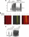
FIGURE 3.
Ataxin-3 preferentially hydrolyzes newly added Ub conjugates on parkin. A, ataxin-3 deubiquitinates newly added Ub conjugates on parkin. In vitro ubiquitination reactions were carried out with GST-parkin bound to glutathione beads at 37 °C. After 1 h, ataxin-3 was added to ongoing reactions (lanes 7–9) and compared with reactions lacking a DUB (lanes 4–6) 1, 2, and 3 h after the addition of ataxin-3. A reaction containing inactive ataxin-3C14S was also added for 3 h as a control. Reactions were immunoblotted for Ub. B, ataxin-3 edits Ub conjugates in real-time as they form on parkin. In vitro ubiquitination reactions were carried out with GST-parkin bound to glutathione beads for 1 h at 37 °C with HA-Ub, washed, and added into a second reaction containing Myc-Ub in the presence or absence of His-ataxin-3 (14Q). Reactions were analyzed using the two-color (HA, red, and Myc, green) LI-COR Odyssey infrared imaging system. C, ataxin-3 edits growing Ub chains on parkin in real-time. In vitro ubiquitination assays with eluted MBP-parkin and FLAG-Ub were incubated for 1 h at 37 °C. After 1 h, ubiquitinated MBP-parkin was immunoprecipitated with FLAG resin, eluted, and added into a second reaction incubated for 1 h at 37 °C containing HA-Ub in the presence or absence of His-ataxin-3 (14Q) or His-ataxin-3C14S. Reactions were immunoblotted for FLAG and HA. MBP-parkin is the band found above 82 kDa. WB, Western blot.
FIGURE 4.
FIGURE 4.
Ataxin-3 interacts with the E2-conjugating enzyme. A, ataxin-3 interacts directly with Ubc7. Wild-type His-ataxin-3 (14Q) or inactive ataxin-3C14S was incubated either alone or in the presence of His-Ubc7 and the cross-linker DTSSP for 1 h at RT. Reactions were terminated following the addition of 1 m Tris, pH 7.5. Samples were boiled in the presence or absence of DTT and immunoblotted for Atx3 and His. B, ataxin-3 binds to UbcH7. Wild-type His-ataxin-3 (14Q) or inactive ataxin-3C14S were incubated either alone or in the presence of UbcH7 and the cross-linker DTSSP for 1 h at RT. Reactions were immunoblotted for His. C, ataxin-3 can interact with the inactive C86S mutant UbcH7. Wild-type His-ataxin-3 (14Q) or inactive ataxin-3C14S were incubated either alone or in the presence of wild-type or the inactive C86S mutant form of UbcH7 and the cross-linker DTSSP for 1 h at RT. Reactions were immunoblotted for His. WB, Western blot.
FIGURE 5.
FIGURE 5.
Initial cycles of E2·Ub thioester complex formation is impeded by ataxin-3 in a parkin-dependent manner. A, ataxin-3 prevents the formation of the E2·Ub thioester complex in a parkin-dependent manner. His-Ubc7 was incubated in the presence of E1, Ub, ATP, and Mg2+ at 37 °C for 4 min. To the indicated reactions, either His-ataxin-3 (14Q) or His-ataxin-3C14S were added in the absence or presence of increased concentrations of MBP-parkin. After 4 min, reactions were terminated by adding 3× SDS sample buffer (+/−DTT). Reactions were immunoblotted for His. B, charging of UbcH7. UbcH7 was incubated in the presence of E1, Ub, ATP, and Mg2+ at 37 °C for the indicated time intervals. Reactions were terminated by adding 3× SDS sample buffer (+/−DTT), followed by staining with Coomassie Blue. C, ataxin-3 opposes UbcH7 charging in a parkin-dependent manner. UbcH7 was charged as described in B for 3 min at 37 °C. To the indicated reactions, either His-ataxin-3 (14Q) or His-ataxin-3C14S was added in the absence or presence of MBP-parkin. Reactions were terminated by adding 3× SDS sample buffer (+/−DTT), followed by staining with Coomassie Blue. Note: the presence of tris(2-carboxyethyl)phosphine instead of DTT allowed for efficient charging of UbcH7. However, its presence made it difficult to discern a clear band for MBP-parkin. WB, Western blot.
FIGURE 6.
FIGURE 6.
Parkin-bound ataxin-3 impedes dissociation of the bound E2-conjugating enzyme. A, parkin co-immunoprecipitates with ataxin-3 in cells. SH-SY5 cells were lysed and immunoprecipitated with either rabbit IgG serum or the rabbit ataxin-3 polyclonal, followed by immunoblotting for ataxin-3 and parkin. B, parkin co-immunoprecipitates with ataxin-3 in mice brain synaptosomal fractions. Synaptosomal fractions were isolated from the brains of parkin wild-type or knock-out mice as described previously (26). Fractions were immunoprecipitated with either rabbit IgG serum or the rabbit ataxin-3 polyclonal antibody, and immunoblotted for ataxin-3 and parkin. C, parkin forms a stable complex with Ubc7 and catalytically active ataxin-3. HEK293 cells were co-transfected with the indicated combinations of FLAG-parkin, Myc-Ubc7, and either wild-type Myc-ataxin-3 (14Q) or the inactive Myc-ataxin-3C14S. Cell lysates were harvested 48 h after transfection and immunoprecipitated with FLAG-resin and immunoblotted for Myc, ataxin-3, Ubc7, and FLAG. WB, Western blot.
FIGURE 7.
FIGURE 7.
Model to explain the functional coupling between ataxin-3-mediated deubiquitination and parkin ubiquitination. When parkin ubiquitinates itself, the charged E2-Ub thioester associates with the RIRparkin, transferring the Ub to a lysine within parkin (1). To form a new thioester conjugate, the uncharged E2 must dissociate from parkin (2), allowing it to interact with the E1-Ub thioester. Binding to the E1-Ub results in the formation of a new E2-Ub (3) that is now free to associate with and ubiquitinate parkin (4). When ataxin-3 is present, it can also interact with the E2-Ub and redirect the transfer of Ub from the E2 away from parkin and onto itself (5). As ataxin-3 dramatically stabilizes the interaction between parkin and the E2, it reduces the ability of the E2 to dissociate from parkin, thus decreasing the efficiency of E2 recharging by the E1 (6). Moreover, the E2 stably bound to parkin also prevents binding with a subsequent charged E2-Ub thioester (7).

References

    1. Kerscher O., Felberbaum R., Hochstrasser M. (2006) Annu. Rev. Cell Dev. Biol. 22, 159–180 - PubMed
    1. Pickart C. M., Eddins M. J. (2004) Biochim. Biophys. Acta 1695, 55–72 - PubMed
    1. Pelzer C., Kassner I., Matentzoglu K., Singh R. K., Wollscheid H. P., Scheffner M., Schmidtke G., Groettrup M. (2007) J. Biol. Chem. 282, 23010–23014 - PubMed
    1. Groettrup M., Pelzer C., Schmidtke G., Hofmann K. (2008) Trends Biochem. Sci. 33, 230–237 - PubMed
    1. Ye Y., Rape M. (2009) Nat. Rev. Mol. Cell Biol. 10, 755–764 - PMC - PubMed

Publication types

LinkOut - more resources