. 2011 Oct 1;15(5):177-182.
doi: 10.1016/j.cossms.2011.05.002.

Polymer physics of the cytoskeleton

Affiliations

Polymer physics of the cytoskeleton

Qi Wen et al. Curr Opin Solid State Mater Sci. .

Abstract

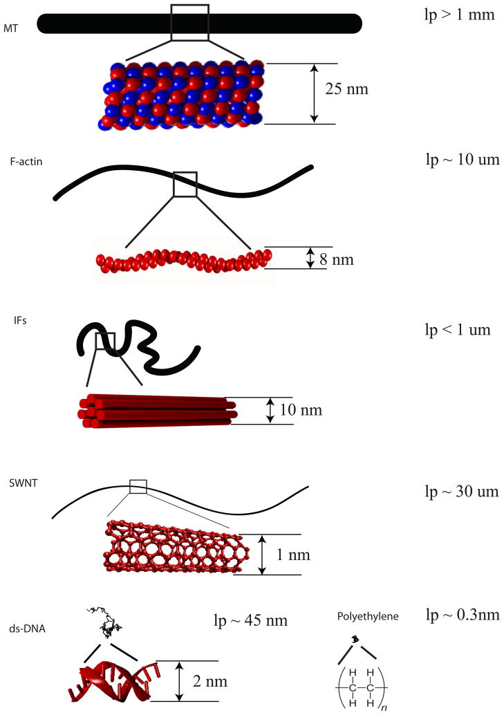
The cytoskeleton is generally visualized by light or electron microscopy as a meshwork of protein filaments that spans the space between the nuclear envelope and the plasma membrane. In most cell types, this meshwork is formed by a three dimensional composite network of actin filaments, microtubules (MT), and intermediate filaments (IF) together with the host of proteins that bind to the sides or ends of these linear polymers. Cytoskeletal binding proteins regulate filament length, crosslink filaments to each other, and apply forces to the filaments. One approach to modeling the mechanical properties of the cytoskeleton and of cell in general is to consider the elements of the cytoskeleton as polymers, using experimental methods and theoretical models developed for traditional polymers but modified for the much larger, stiffer, and fragile biopolymers comprising the cytoskeleton. The presence of motor proteins that move actin filaments and microtubules also creates a new class of active materials that are out of thermodynamic equilibrium, and unconstrained by limitations of the fluctuation-dissipation theorem. These active materials create rich opportunities for experimental design and theoretical developments. The degree to which the mechanics of live cells can usefully be modeled as highly complex polymer networks is by no means certain, and this article will discuss recent progress in quantitatively measuring cytoskeletal polymer systems and relating them to the properties of the cell.


Figures

Figure 1
Figure 1. Elongated structure of cytoskeletal filaments
Microtubules, F-actin, and intermediate filaments with contour length 10 um are drawn at the same scale in comparison to single wall carbon nanotubes, double strand DNA, and polyethylene of the same contour length.
Figure 2
Figure 2. Effect of actin binding proteins and molecular motors on rheological properties of cytoskeletal networks
(A) Strain stiffening in isotropic semiflexible polymer networks (adapted from [45] ) due to the nonlinear force extension of individual filaments. (B) Flexible cross-linkers in the actin network lead to strain stiffening of the network (adapted from [37]). (C) Myosin motors in the crosslinked actin network generate internal tension that stiffen the network (adapted from [25]).
Figure 3
Figure 3. Surface charge density of cytoskeletal filaments
The charge density of F-actin, microtubules and two types of IFs Surface charge density was calculated from the amino acid sequence for the human variants of these proteins derived from GenBank without regard to post-translational modifications, except for actin, where amino acid processing and binding of charged ligands contribute a significant amount of charge to the protein. The charge per length of IFs is calculated from the amino acid sequence of IF subunits before post-translational modifications and the assumptions that all IFs contain 32 monomers in the cross-section of a 10 nm cylindrical filament . The molar ratio of neurofilament triplet proteins is taken as NFH:NFM:NFL = 1:2:6. The value for actin is for post-translationally modified beta-actin, the most prominent non-muscle cytoplasmic actin isoform assuming 370 monomers per micron of 8 nm filament and that each monomer binds 1 Mg2+ and 1 ADP3-. Microtubule charge assumes 1625 tubulin dimers per micron of 25 nm diameter MT, with each dimer binding 2 Mg2+ and 2 GDP3-. The calculated charge per length of the filament was divided by the diameter of each filament type.

References

    1. Fletcher DA, Mullins RD. Cell mechanics and the cytoskeleton. Nature. 2010;463:485–92. A beautiful review of recent progresses in understanding the role of cytoskeleton in the mechanics of cells. - PMC - PubMed
    1. Pelling AE, Horton MA. An historical perspective on cell mechanics. Pflugers Arch. 2008;456:3–12. - PubMed
    1. Zhou EH, Trepat X, Park CY, Lenormand G, Oliver MN, Mijailovich SM, et al. Universal behavior of the osmotically compressed cell and its analogy to the colloidal glass transition. Proc Natl Acad Sci U S A. 2009;106:10632–7. - PMC - PubMed
    1. Mofrad MRK. Rheology of the Cytoskeleton. Annual Review of Fluid Mechanics. 2009;41:433–53. A comprehensive review of the key experiments and theories describing rheology and mechanics of the cytoskeleton.
    1. Sept D, MacKintosh FC. Microtubule elasticity: connecting all-atom simulations with continuum mechanics. Phys Rev Lett. 2010;104:018101. Pioneering work that begins to link molecular level simulations to whole filament properties. - PubMed

LinkOut - more resources