

Antimicrobial properties of amyloid peptides
- PMID: 22081976
- PMCID: PMC3297685
- DOI: 10.1021/mp200419b
Antimicrobial properties of amyloid peptides
Abstract
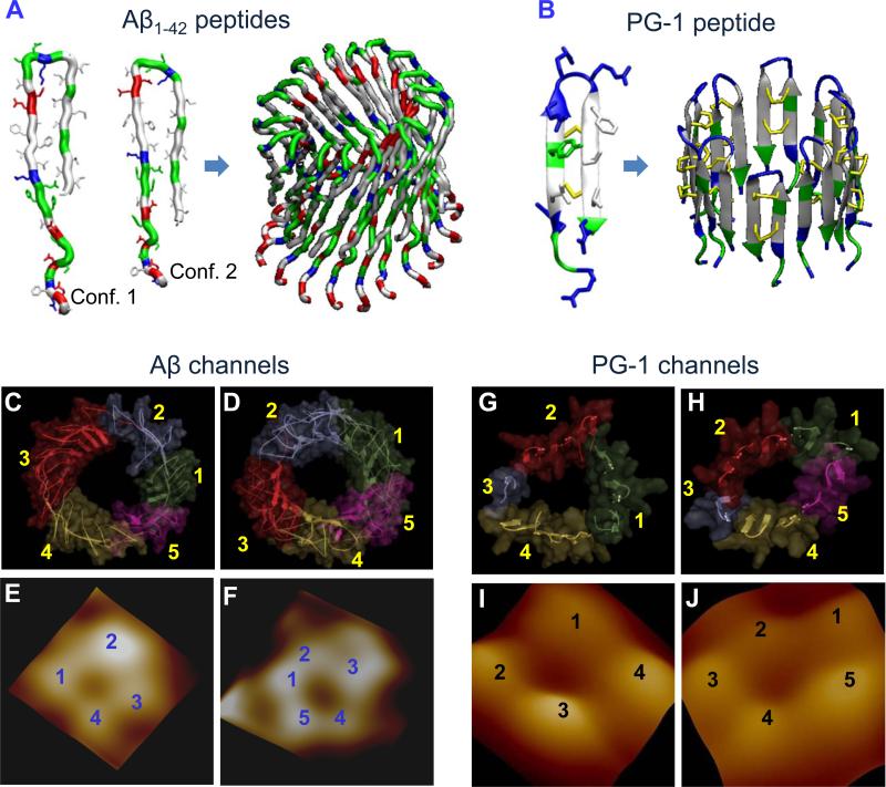
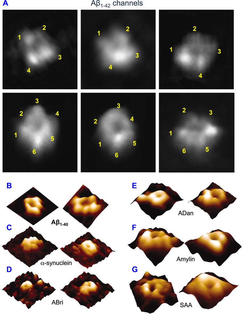
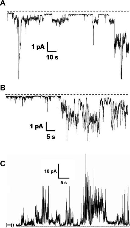
More than two dozen clinical syndromes known as amyloid diseases are characterized by the buildup of extended insoluble fibrillar deposits in tissues. These amorphous Congo red staining deposits known as amyloids exhibit a characteristic green birefringence and cross-β structure. Substantial evidence implicates oligomeric intermediates of amyloids as toxic species in the pathogenesis of these chronic disease states. A growing body of data has suggested that these toxic species form ion channels in cellular membranes causing disruption of calcium homeostasis, membrane depolarization, energy drainage, and in some cases apoptosis. Amyloid peptide channels exhibit a number of common biological properties including the universal U-shape β-strand-turn-β-strand structure, irreversible and spontaneous insertion into membranes, production of large heterogeneous single-channel conductances, relatively poor ion selectivity, inhibition by Congo red, and channel blockade by zinc. Recent evidence has suggested that increased amounts of amyloids not only are toxic to its host target cells but also possess antimicrobial activity. Furthermore, at least one human antimicrobial peptide, protegrin-1, which kills microbes by a channel-forming mechanism, has been shown to possess the ability to form extended amyloid fibrils very similar to those of classic disease-forming amyloids. In this paper, we will review the reported antimicrobial properties of amyloids and the implications of these discoveries for our understanding of amyloid structure and function.
Figures
References
-
- Cohen AS. General introduction and a brief history of the amyloid fibril. Nijhoff; Dordrecht: 1986. pp. 3–19.
-
- Hirakura Y, Lin MC, Kagan BL. Alzheimer amyloid Aβ1-42 channels: effects of solvent, pH, and Congo Red. J Neurosci Res. 1999;57:458–66. - PubMed
-
- Gertz MA. The classification and typing of amyloid deposits. Am J Clin Pathol. 2004;121:787–9. - PubMed
-
- Sunde M, Serpell LC, Bartlam M, Fraser PE, Pepys MB, Blake CC. Common core structure of amyloid fibrils by synchrotron X-ray diffraction. J Mol Biol. 1997;273:729–39. - PubMed
Publication types
MeSH terms
Substances
Grants and funding
LinkOut - more resources
Full Text Sources
Other Literature Sources
