

Multiscale photoacoustic microscopy of single-walled carbon nanotube-incorporated tissue engineering scaffolds
- PMID: 22082018
- PMCID: PMC3311878
- DOI: 10.1089/ten.TEC.2011.0519
Multiscale photoacoustic microscopy of single-walled carbon nanotube-incorporated tissue engineering scaffolds
Abstract
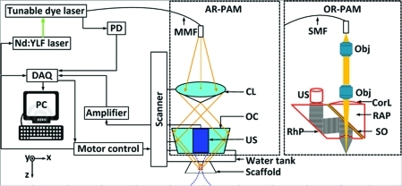
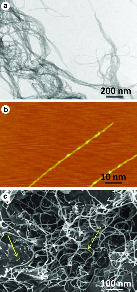
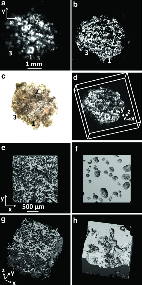
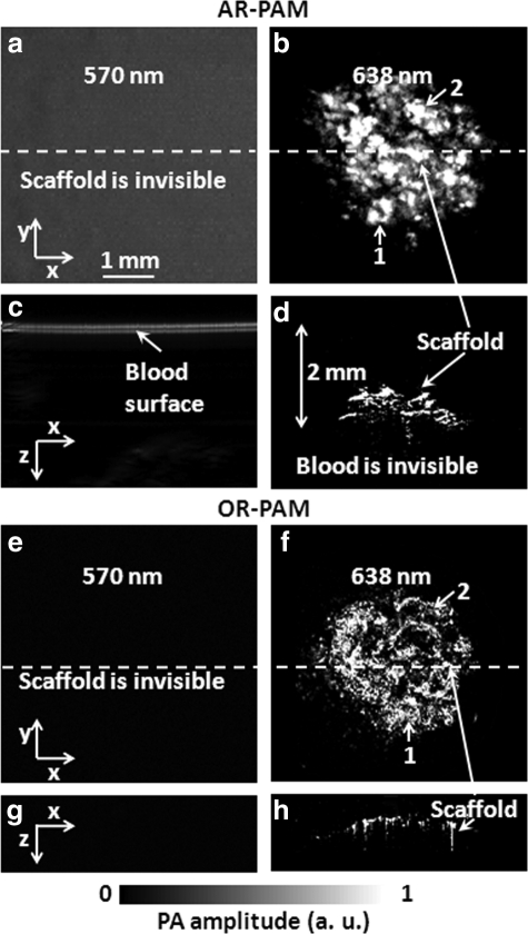
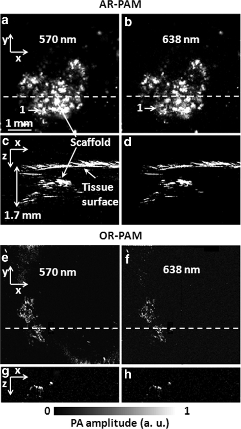
Three-dimensional polymeric scaffolds provide structural support and function as substrates for cells and bioactive molecules necessary for tissue regeneration. Noninvasive real-time imaging of scaffolds and/or the process of tissue formation within the scaffold remains a challenge. Microcomputed tomography, the widely used technique to characterize polymeric scaffolds, shows poor contrast for scaffolds immersed in biological fluids, thereby limiting its utilities under physiological conditions. In this article, multiscale photoacoustic microscopy (PAM), consisting of both acoustic-resolution PAM (AR-PAM) and optical-resolution PAM (OR-PAM), was employed to image and characterize single-walled carbon-nanotube (SWNT)-incorporated poly(lactic-co-glycolic acid) polymer scaffolds immersed in biological buffer. SWNTs were incorporated to reinforce the mechanical properties of the scaffolds, and to enhance the photoacoustic signal from the scaffolds. By choosing excitation wavelengths of 570 and 638 nm, multiscale PAM could spectroscopically differentiate the photoacoustic signals generated from blood and from carbon-nanotube-incorporated scaffolds. OR-PAM, providing a fine lateral resolution of 2.6 μm with an adequate tissue penetration of 660 μm, successfully quantified the average porosity and pore size of the scaffolds to be 86.5%±1.2% and 153±15 μm in diameter, respectively. AR-PAM further extended the tissue penetration to 2 mm at the expense of lateral resolution (45 μm). Our results suggest that PAM is a promising tool for noninvasive real-time imaging and monitoring of tissue engineering scaffolds in vitro, and in vivo under physiological conditions.
Figures
Similar articles
-
Multimodal ultrasound-photoacoustic imaging of tissue engineering scaffolds and blood oxygen saturation in and around the scaffolds.Tissue Eng Part C Methods. 2014 May;20(5):440-9. doi: 10.1089/ten.TEC.2013.0203. Epub 2014 Feb 28. Tissue Eng Part C Methods. 2014. PMID: 24107069 Free PMC article.
-
Investigation of neovascularization in three-dimensional porous scaffolds in vivo by a combination of multiscale photoacoustic microscopy and optical coherence tomography.Tissue Eng Part C Methods. 2013 Mar;19(3):196-204. doi: 10.1089/ten.TEC.2012.0326. Epub 2012 Sep 7. Tissue Eng Part C Methods. 2013. PMID: 22838500 Free PMC article.
-
Functionalized carbon nanotube reinforced scaffolds for bone regenerative engineering: fabrication, in vitro and in vivo evaluation.Biomed Mater. 2014 Jun;9(3):035001. doi: 10.1088/1748-6041/9/3/035001. Epub 2014 Mar 31. Biomed Mater. 2014. PMID: 24687391
-
Performance comparison of PLA- and PLGA-coated porous bioceramic scaffolds: Mechanical, biodegradability, bioactivity, delivery and biocompatibility assessments.J Control Release. 2022 Nov;351:1-7. doi: 10.1016/j.jconrel.2022.09.022. Epub 2022 Sep 18. J Control Release. 2022. PMID: 36115555 Review.
-
The paths of musculoskeletal scaffold research leading to long-term effects.J Long Term Eff Med Implants. 2012;22(3):237-342. doi: 10.1615/jlongtermeffmedimplants.2013006182. J Long Term Eff Med Implants. 2012. PMID: 23582115 Review.
Cited by
-
Towards non-contact photoacoustic imaging [review].Photoacoustics. 2020 Sep 23;20:100207. doi: 10.1016/j.pacs.2020.100207. eCollection 2020 Dec. Photoacoustics. 2020. PMID: 33024694 Free PMC article. Review.
-
Characterization of mechanical and biological properties of 3-D scaffolds reinforced with zinc oxide for bone tissue engineering.PLoS One. 2014 Jan 31;9(1):e87755. doi: 10.1371/journal.pone.0087755. eCollection 2014. PLoS One. 2014. PMID: 24498185 Free PMC article.
-
Photoacoustic Imaging in Tissue Engineering and Regenerative Medicine.Tissue Eng Part B Rev. 2020 Feb;26(1):79-102. doi: 10.1089/ten.TEB.2019.0296. Epub 2020 Jan 14. Tissue Eng Part B Rev. 2020. PMID: 31854242 Free PMC article. Review.
-
In Vivo Tracking of Tissue Engineered Constructs.Micromachines (Basel). 2019 Jul 16;10(7):474. doi: 10.3390/mi10070474. Micromachines (Basel). 2019. PMID: 31315207 Free PMC article. Review.
-
Monitoring/Imaging and Regenerative Agents for Enhancing Tissue Engineering Characterization and Therapies.Ann Biomed Eng. 2016 Mar;44(3):750-72. doi: 10.1007/s10439-015-1509-y. Epub 2015 Dec 21. Ann Biomed Eng. 2016. PMID: 26692081 Free PMC article. Review.
References
-
- Karageorgiou V. Kaplan D. Porosity of 3D biornaterial scaffolds and osteogenesis. Biomaterials. 2005;26:5474. - PubMed
Publication types
MeSH terms
Substances
Grants and funding
LinkOut - more resources
Full Text Sources
Research Materials
Miscellaneous
