Developmental aspects of the intracerebral microvasculature and perivascular spaces: insights into brain response to late-life diseases
- PMID: 22082663
- PMCID: PMC3227674
- DOI: 10.1097/NEN.0b013e31823ac627
Developmental aspects of the intracerebral microvasculature and perivascular spaces: insights into brain response to late-life diseases
Abstract
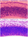
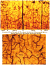
The development of the microvasculature of the human cerebral cortex offers insight into the response of the cerebral cortex to later-life brain injury. We describe the 3 basic and distinct components of the developmental anatomy of the cerebral cortical microvascular system. The first compartment is meningeal and, therefore, extracerebral. In addition to the major venous sinuses, arachnoidal arteries, and veins, the pial anastomotic capillary plexus that covers the surface of the developing and adult cerebral cortex represents the source of thepenetrating vessels that become the second component, the intracerebral extrinsic microvascular compartment. During embryogenesis, sprouting vascular elements from pial capillaries pierce the brain's external glial limiting membrane and penetrate the cortex. These vessels, which eventually differentiate into arterioles and venules, are separated from the cortical tissue by the extravascular Virchow-Robin compartment (V-RC) formed between the internal vascular and the external glial basal laminae. The V-RC remains open to the meningeal interstitial spaces and outside the blood-brain barrier (BBB) and acts asa prelymphatic drainage system for removal of substances that cannot be transported into the blood or catabolized intracellularly. The third element is the dense intracerebralintrinsic microvascular compartment. Intracerebral capillary vessels sprout from the perforating vessels, penetrate through the Virchow-Robin glial membrane, and enter the neuropil. Intracerebral capillaries lack smooth muscle and a V-RC and consist only of endothelial cells separated from the intracerebral space by a basal lamina. Their role as the physiological BBB is the exchange of oxygen, glucose, and small molecules. This developmental perspective highlights 3 principles: (a) the V-RC is intimately related to the cortical penetrating arterioles and venules and represents an inefficient protolymphatic system that lacks the anatomic and physiological constituents found in lymphatic beds elsewhere in the body; (b)the anatomic contiguity of the V-RC and the penetrating vascular compartment (arterioles and venules) implies that the pathology in 1 compartment could lead to dysfunction in the others; and (c) the anatomic localization of the immunologic BBB at the level of the penetrating venules might impose constraints on immunologically mediated transport involving the V-RC.
Figures


References
-
- White L, Petrovitch H, Hardman J, et al. Cerebrovascular pathology and dementia in autopsied Honolulu-Asia Aging Study participants. Ann N Y Acad Sci. 2002;977:9–23. - PubMed
-
- Kalaria RN, Kenny RA, Ballard CG, et al. Towards defining the neuropathological substrates of vascular dementia. J Neurol Sci. 2004;226:75–80. - PubMed
-
- Vinters HV, Ellis WG, Zarow C, et al. Neuropathologic substrates of ischemic vascular dementia. J Neuropathol Exp Neurol. 2000;59:931–45. - PubMed
-
- Vinters HV, Wang ZZ, Secor DL. Brain parenchymal and microvascular amyloid in Alzheimer’s disease. Brain Pathol. 1996;6:179–95. - PubMed
Publication types
MeSH terms
Grants and funding
LinkOut - more resources
Full Text Sources
Other Literature Sources

