. 2011 Dec;7(12):1448-61.
doi: 10.4161/auto.7.12.17802.

Resveratrol-mediated autophagy requires WIPI-1-regulated LC3 lipidation in the absence of induced phagophore formation

Affiliations

Resveratrol-mediated autophagy requires WIPI-1-regulated LC3 lipidation in the absence of induced phagophore formation

Mario Mauthe et al. Autophagy. 2011 Dec.

Abstract

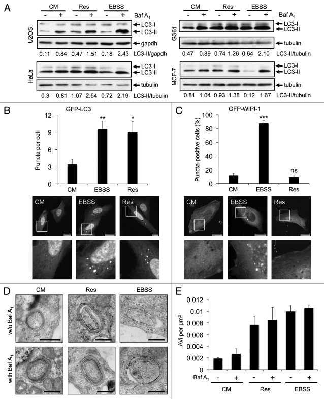
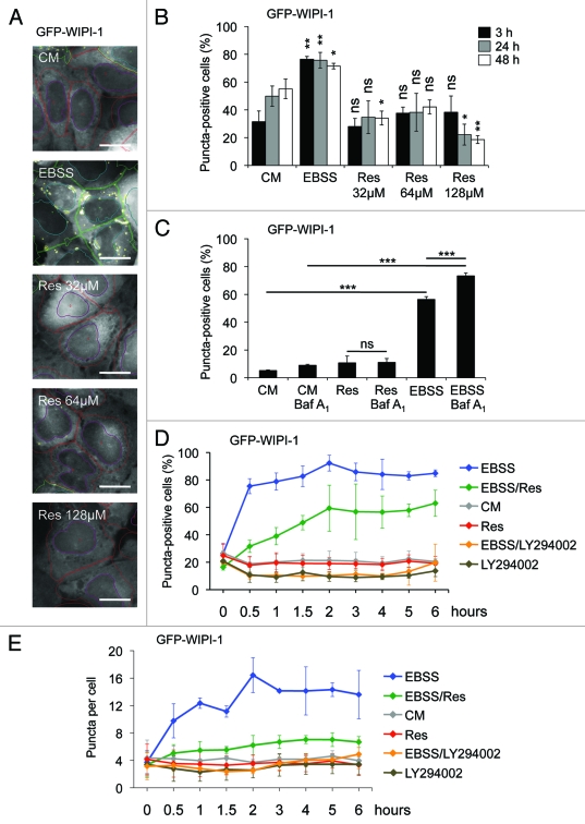
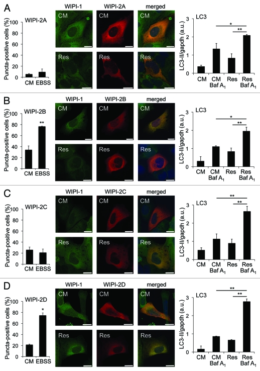
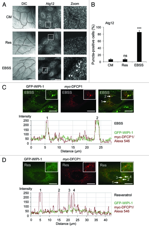
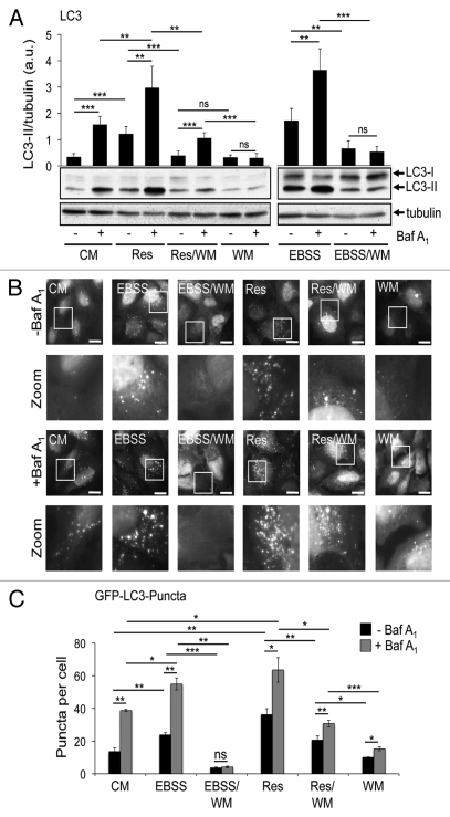
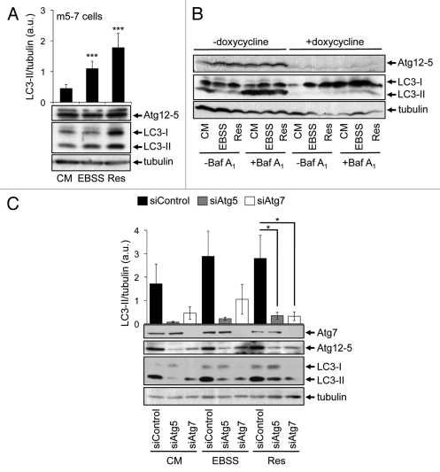
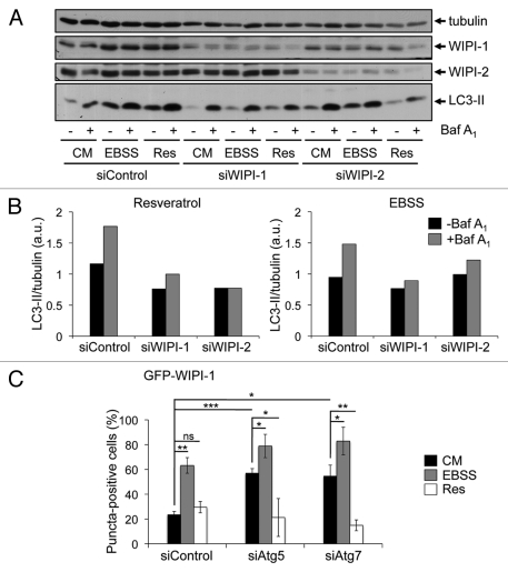
Canonical autophagy is positively regulated by the Beclin 1/phosphatidylinositol 3-kinase class III (PtdIns3KC3) complex that generates an essential phospholipid, phosphatidylinositol 3-phosphate (PtdIns(3)P), for the formation of autophagosomes. Previously, we identified the human WIPI protein family and found that WIPI-1 specifically binds PtdIns(3)P, accumulates at the phagophore and becomes a membrane protein of generated autophagosomes. Combining siRNA-mediated protein downregulation with automated high through-put analysis of PtdIns(3)P-dependent autophagosomal membrane localization of WIPI-1, we found that WIPI-1 functions upstream of both Atg7 and Atg5, and stimulates an increase of LC3-II upon nutrient starvation. Resveratrol-mediated autophagy was shown to enter autophagic degradation in a noncanonical manner, independent of Beclin 1 but dependent on Atg7 and Atg5. By using electron microscopy, LC3 lipidation and GFP-LC3 puncta-formation assays we confirmed these results and found that this effect is partially wortmannin-insensitive. In line with this, resveratrol did not promote phagophore localization of WIPI-1, WIPI-2 or the Atg16L complex above basal level. In fact, the presence of resveratrol in nutrient-free conditions inhibited phagophore localization of WIPI-1. Nevertheless, we found that resveratrol-mediated autophagy functionally depends on canonical-driven LC3-II production, as shown by siRNA-mediated downregulation of WIPI-1 or WIPI-2. From this it is tempting to speculate that resveratrol promotes noncanonical autophagic degradation downstream of the PtdIns(3)P-WIPI-Atg7-Atg5 pathway, by engaging a distinct subset of LC3-II that might be generated at membrane origins apart from canonical phagophore structures.


Figures

Figure 1.
Figure 1.
Resveratrol promotes an increase of autophagosomes. U2OS, HeLa, G361 and MCF-7 cells were treated with 64 µM resveratrol (Res), nutrient-free medium (EBSS) or control medium (CM) for 3 h. Bafilomycin A1 (Baf A1) was added after 2 h as indicated (+). LC3-II protein abundance was determined and normalized over tubulin or gapdh. The quantification of one representative experiment (n = 3 for each cell line) is shown (A). U2OS cells were transiently transfected with GFP-LC3 (B) or GFP-WIPI-1 (C) and treated with 64 µM resveratrol (Res), nutrient-free medium (EBSS) or control medium (CM) for 3 h and images were acquired by confocal microscopy. The number of GFP-LC3 puncta per cell was determined from 33–36 individual cells (n = 3) by using Z-stack projections and ImagePro Plus 4.1 analysis software (B, upper panel). The number of GFP-WIPI-1 puncta-positive cells per treatment was determined from 400 individual cells (n = 4) by fluorescent microscopy (C, upper panel). The results were expressed as mean ± SD p-values: *p < 0.05; **p < 0.01; ***p < 0.001; ns: not significant. Scale bars: 20 µm. Stable GFP-WIPI-1 U2OS cells were treated with control medium (CM), 64 µM resveratrol (Res) or nutrient-free medium (EBSS) in the presence (+) or absence (-) of bafilomycin A1 (Baf A1). For EM analysis, ultrathin resin sections of different samples were used and representative images are shown (D). Scale bars: 400 nm. The number of multilayered autophagosomal vesicular structures (AVi) per square micrometer was determined for each treatment (E).
Figure 2.
Figure 2.
WIPI-1 puncta formation is stimulated upon nutrient starvation but not upon resveratrol treatment. Stable GFP-WIPI-1 U2OS cells were treated with control medium (CM), 32 μM, 64 μM or 128 μM resveratrol (Res) or nutrient-free medium (EBSS) for 3, 24 or 48 h. GFP-WIPI-1 fluorescence images were automatically acquired and analyzed (A). The number of GFP-WIPI-1 puncta-positive cells was determined and each bar represents mean values of 1732–3899 individually analyzed cells (n = 3) ± SD (B). Stable GFP-WIPI-1 U2OS cells were treated with control medium (CM) or 64 μM resveratrol (Res) in the presence or absence of bafilomycin A1 (Baf A1) or nutrient-free medium (EBSS) for 3 h. Automated GFP-WIPI-1 puncta-formation analysis was conducted (n = 5, 16044–22479 individually analyzed cells) and expressed as the number of GFP-WIPI-1 puncta-positive cells ± SD (C). A time-course experiment was conducted by using the GFP-WIPI-1 U2OS cell line and the following treatments: control medium (CM), 64 μM resveratrol (Res), nutrient-free medium (EBSS), 100 μM LY294002 in control medium (LY294002), 64 μM resveratrol in nutrient-free medium (EBSS/Res) or 100 μM LY294002 in nutrient-free medium (EBSS/LY294002). Automated GFP-WIPI-1 puncta-formation analysis from up to 5000 individual cells per treatment (n = 3) was expressed as the number of GFP-WIPI-1 puncta-positive cells ± SD (D) or the number of GFP-WIPI-1 puncta per cell (E). p-values: *p < 0.05; **p < 0.01; ***p < 0.001; ns = not significant. Scale bars: 20 µm.
Figure 3.
Figure 3.
WIPI-2 puncta formation is not induced upon resveratrol treatment, but WIPI-2B and WIPI-2D form puncta upon nutrient starvation. U2OS cells were transiently tranfected with either of four GFP-WIPI-2 variants (GFP-WIPI-2A, -2B, -2C, -2D) and treated with control medium (CM) or nutrient-free medium (EBSS). The number of GFP-WIPI-2 puncta-positive cells was determined by fluorescence microscopy and mean values from 400–600 cells (n = 2–3) are presented ± SD (A–D, left panels). Stable GFP-WIPI-1 U2OS cells were transiently transfected with myc-tagged WIPI-2 (myc-WIPI-2A, -2B, -2C, -2D), treated with or without 64 µM resveratrol (Res) in control medium (CM) for 3 h followed by anti-myc/Alexa546 immunostaining and confocal microscopy (representative images, n = 2). Nuclei were stained with TO-PRO-3 (A–D, middle panels). In parallel, U2OS cells coexpressing myc-tagged WIPI-2 variants (myc-WIPI-2A, -2B, -2C, -2D) and GFP-WIPI-1, treated with or without 64 µM resveratrol (Res) in control medium (CM) for 3 h in the presence or absence of bafilomycin A1 (Baf A1) were subjected to LC3-II protein monitoring (A–D, right panels). Each bar represents mean values of three independent experiments ± SD p-values: *p < 0.05; **p < 0.01. Scale bars: 20 µm.
Figure 4.
Figure 4.
Increase of Atg12 puncta and WIPI-1/DFCP1 colocalization upon nutrient-starvation but not upon resveratrol treatment. U2OS cells were treated with 64 µM resveratrol (Res), nutrient-free medium (EBSS) or control medium (CM) for 3 h, followed by anti-Atg12/Alexa488 immunostaining in order to visualize the endogenous Atg16L complex by confocal microscopy (A). The number of Atg12 puncta-positive cells was determined (600 cells, n = 3) and each bar represents mean values ± SD (B). p-values: ***p < 0.001; ns = not significant. Stable GFP-WIPI-1 U2OS cells were transiently transfected with myc-DFCP1, treated with nutrient-free medium (EBSS) or with 64 µM resveratrol (Res) for 3 h followed by anti-myc/Alexa546 immunostaining. Representative confocal microscopy images (n = 3) from individual optical sections (upper panels) along with the corresponding intensity profiles (lower panels) are presented (C and D). Scale bars: 20 µm.
Figure 5.
Figure 5.
Resveratrol-promoted increase of LC3-II protein abundance and GFP-LC3 puncta is partially insensitive to wortmannin treatment. Stable GFP-WIPI-1 U2OS cells were treated with control medium (CM), resveratrol (Res), wortmannin (WM), resveratrol plus wortmannin (Res/WM), nutrient-free medium (EBSS) or nutrient-free medium plus wortmannin (EBSS/WM) for 3 h. Bafilomycin A1 (Baf A1) was added after 2 h as indicated (+). LC3-II protein abundance was determined and normalized over tubulin. Each bar represents mean values ± SD (n = 6) (A). Stable GFP-LC3 U2OS cells were treated as above and subjected to automated fluorescence image acquisition (B) and analysis (C). Each bar represents mean values ± SD from the analysis of 1553–3207 individual cells (n = 3). p-values: *p < 0.05; **p < 0.01; ***p < 0.001; ns: not significant. Scale bars: 20 µm.
Figure 6.
Figure 6.
Resveratrol-mediated autophagy depends on Atg7 and Atg5. Mouse embryonic fibroblasts (m5-7 cells) were treated with 64 µM resveratrol (Res), nutrient-free medium (EBSS) or control medium (CM) for 3 h and subjected to quantitative anti-LC3 western blotting (n = 7) (A). Doxycycline (1 µg/ml) was added to the culture medium in order to shut down Atg5 expression in m5-7 cells. Representative western blots (n = 3) detecting Atg5 (Atg12–5 conjugate), LC3 or tubulin upon treatments with control medium (CM), 64 µM resveratrol (Res) or nutrient-free medium (EBSS) for 3 h are shown; bafilomycin A1 (Baf A1) was added after 2 h as indicated (B). Stable GFP-WIPI-1 U2OS cells were transfected with control, Atg5 or Atg7 siRNAs for 72 h and treated with control medium (CM), 64 µM resveratrol (Res) or nutrient-free medium (EBSS) for 3 h. Representative downregulation of Atg5 (Atg12–5 conjugate) or Atg7 protein is shown and LC3-II protein abundance was normalized over tubulin and quantified (n = 3) (C). p-values: *p < 0.05; ***p < 0.001.
Figure 7.
Figure 7.
Functional requirement of WIPI-1 and WIPI-2 for LC3 lipidation upon both nutrient starvation and resveratrol treatment. Human G361 cells were transfected with control, WIPI-1 or WIPI-2 siRNA for 72 h and treated with control medium (CM), 64 µM resveratrol (Res) or nutrient-free medium (EBSS) for 3 h. Bafilomycin A1 (Baf A1) was added after 2 h as indicated (+). Representative western blot results (A) along with the quantification of EBSS and resveratrol treatments (B) was shown. Stable GFP-WIPI-1 U2OS cells were transfected with control, Atg5 or Atg7 siRNA for 72 h, treated as above and subjected to automated GFP-WIPI-1 image acquisition and analysis (C). Each bar represents mean values of GFP-WIPI-1 puncta-positive cells (up to 2500 individual cells, n = 3) ± SD p-values: *p < 0.05; **p < 0.01; ***p < 0.001; ns = not significant.

Similar articles

Cited by

References

    1. Mizushima N, Levine B, Cuervo AM, Klionsky DJ. Autophagy fights disease through cellular self-digestion. Nature. 2008;451:1069–75. doi: 10.1038/nature06639. - DOI - PMC - PubMed
    1. Weidberg H, Shvets E, Elazar Z. Biogenesis and Cargo Selectivity of Autophagosomes. Annu Rev Biochem 2010; 10.1146/annurev-biochem-052709-094552 - DOI - PubMed
    1. Klionsky DJ, Cregg JM, Dunn WA, Jr., Emr SD, Sakai Y, Sandoval IV, et al. A unified nomenclature for yeast autophagy-related genes. Dev Cell. 2003;5:539–45. doi: 10.1016/S1534-5807(03)00296-X. - DOI - PubMed
    1. Kawamata T, Kamada Y, Kabeya Y, Sekito T, Ohsumi Y. Organization of the pre-autophagosomal structure responsible for autophagosome formation. Mol Biol Cell. 2008;19:2039–50. doi: 10.1091/mbc.E07-10-1048. - DOI - PMC - PubMed
    1. Ohsumi Y, Mizushima N. Two ubiquitin-like conjugation systems essential for autophagy. Semin Cell Dev Biol. 2004;15:231–6. doi: 10.1016/j.semcdb.2003.12.004. - DOI - PubMed

Publication types

MeSH terms

Substances

LinkOut - more resources