. 2012 Apr;90(4):413-26.
doi: 10.1007/s00109-011-0828-1. Epub 2011 Nov 15.

Suppression of antigen-specific CD4+ T cell activation by SRA/CD204 through reducing the immunostimulatory capability of antigen-presenting cell

Affiliations

Suppression of antigen-specific CD4+ T cell activation by SRA/CD204 through reducing the immunostimulatory capability of antigen-presenting cell

Huanfa Yi et al. J Mol Med (Berl). 2012 Apr.

Abstract

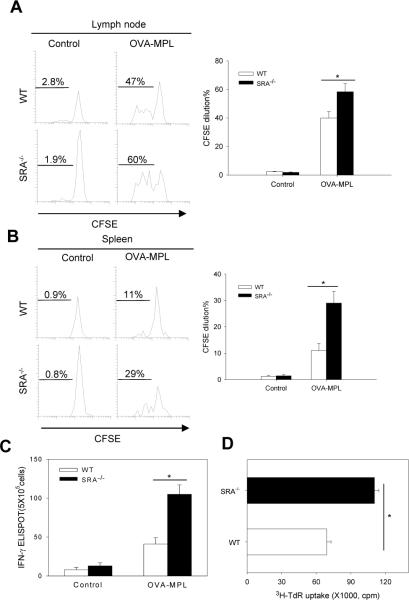
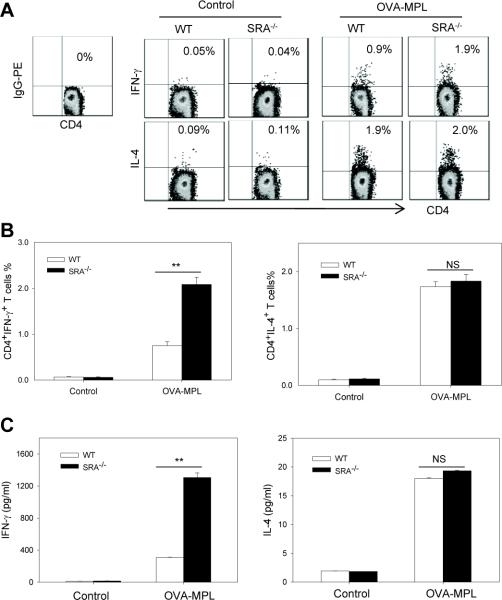
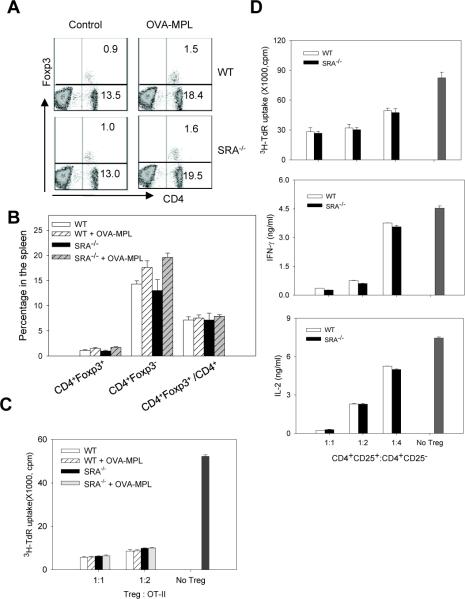
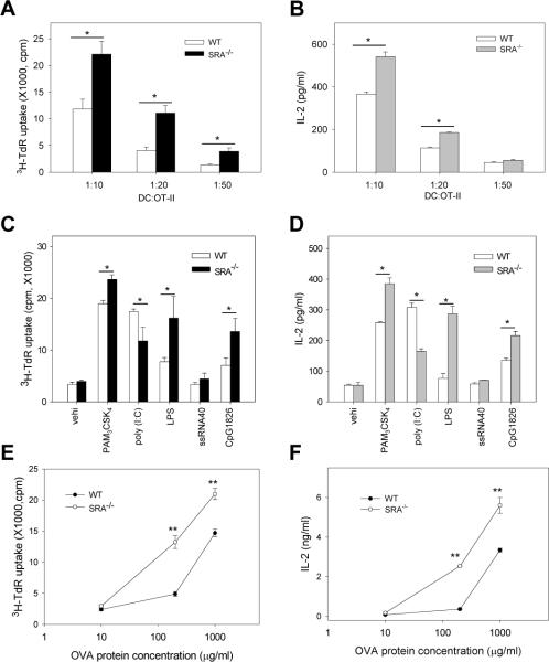
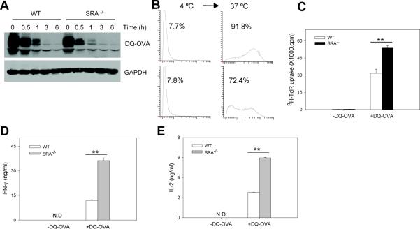
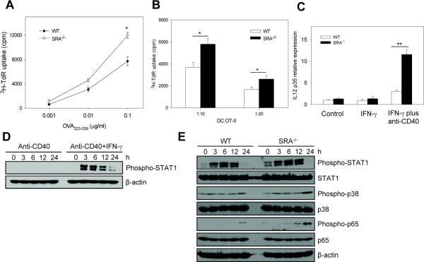
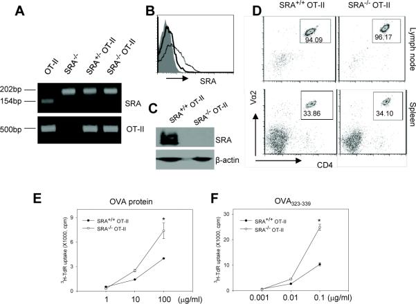
Pattern recognition scavenger receptor SRA/CD204, primarily expressed on specialized antigen-presenting cells (APCs), including dendritic cells (DCs) and macrophages, has been implicated in multiple physiological and pathological processes, including atherosclerosis, Alzheimer's disease, endotoxic shock, host defense, and cancer development. SRA/CD204 was also recently shown to function as an attenuator of vaccine response and antitumor immunity. Here, we, for the first time, report that SRA/CD204 knockout (SRA(-/-)) mice developed a more robust CD4(+) T cell response than wild-type mice after ovalbumin immunization. Splenic DCs from the immunized SRA(-/-) mice were much more efficient than those from WT mice in stimulating naïve OT-II cells, indicating that the suppressive activity of SRA/CD204 is mediated by DCs. Strikingly, antigen-exposed SRA(-/-) DCs with or without lipopolysaccharide treatment exhibited increased T-cell-stimulating activity in vitro, which was independent of the classical endocytic property of the SRA/CD204. Additionally, absence of SRA/CD204 resulted in significantly elevated IL12p35 expression in DCs upon CD40 ligation plus interferon gamma (IFN-γ) stimulation. Molecular studies reveal that SRA/CD204 inhibited the activation of STAT1, mitogen activated protein kinase p38, and nuclear factor-kappa B signaling activation in DCs treated with anti-CD40 antibodies and IFN-γ. Furthermore, splenocytes from the generated SRA(-/-) OT-II mice showed heightened proliferation upon stimulation with OVA protein or MHC-II-restricted OVA(323-339) peptide compared with cells from the SRA(+/+) OT-II mice. These results not only establish a new role of SRA/CD204 in limiting the intrinsic immunogenicity of APCs and CD4(+) T cell activation but also provide additional insights into the molecular mechanisms involved in the immune suppression by this molecule.


Figures

Figure 1
Figure 1. Lack of SRA/CD204 potentiates an OVA-specific CD4+ T cell response by enhancing T cell-stimulating functions of DCs
A and B. SRA/CD204 suppresses OVA-MPL induced expansion of adoptively transferred OT-II cells. Mice (n=5) received CFSE-labeled OT-II cells, followed by OVA-MPL immunization. Draining lymph nodes (A) and spleen (B) were analyzed using FACS by gating on CD4+CSFE+ cells. Representative histograms from two separate experiments with similar results are shown (*p<0.01). C. Increased IFN-γ production by OVA-specific CD4+ T cells from SRA−/− mice. Mice (n=5) were immunized with OVA-MPL or left untreated. Splenocytes were stimulated with OVA323-339 and assayed for IFN-γ secretion using ELISPOT (*p<0.01). D. Increased T cell-stimulating activity of DCs from immunized SRA−/− mice. Splenic CD11c+ DCs were magnetically isolated from the immunized WT or SRA−/− mice and used to stimulate OT-II cells. Cell proliferation was measured using 3H-TdR uptake assays (*p<0.01). Data are means ± SE. Results representative of three independent experiments are shown.
Figure 2
Figure 2. OVA-specific CD4+ T cells from OVA-MPL immunized SRA−/− mice produced more Th1 cytokine IFN-γ
A and B. WT and SRA−/− mice (n=5) were immunized with OVA-MPL twice. Splenocytes were stimulated with OVA323-339 and subjected to intracellular cytokine staining assays. Representative histograms from three independent experiments are shown. B. The effect of SRA/CD204 on the percentage of OVA-specific CD4+IFN-γ+ T cells (left) and CD4+IL-4+ T cells (right) before and after vaccination are also shown (**p<0.005, NS, not significant). C. Supernatants from OVA323-339-stimulated cells were collected and assayed for the levels of IFN-γ (left) and IL-4 (right) by ELISA (**p<0.005). Data are means ± SE and represent three independent experiments.
Figure 3
Figure 3. SRA/CD204 deficiency does not affect Tregs and their suppressive activity
A and B. Following OVA-MPL immunization (n=3), Tregs in the spleen were stained with CD4 and Foxp3 antibodies. Representative histograms from two experiments with similar results are shown (A). The percentage of CD4+Foxp3+ and CD4+Foxp3 cells in total splenocytes, as well as the percentage of CD4+Foxp3+ in total CD4+ cells are also analyzed (B). C. CD4+CD25+ Treg cells were isolated from control or OVA-MPL immunized mice, and co-cultured with OT-II cells. Proliferation of OT-II cells in response to OVA323-339 stimulation was measured using 3H-TdR uptake assays. D. CD4+CD25 cells and CD4+CD25+ Tregs were isolated from OVA-MPL immunized mice, co-cultured at different ratios as indicated. Proliferation of CD4+CD25 effector T cells upon stimulation of CD3/CD28 mAb was examined using 3H-TdR uptake assays (top). IFN-γ (middle) and IL-2 (bottom) levels in the culture supernatants were measured by ELISA. Results representative of two independent experiments with similar results are shown.
Figure 4
Figure 4. SRA/CD204 suppresses the ability of DCs to stimulate OVA-specific CD4+ T cells in vitro in the presence or absence of LPS treatment
A and B. Immunogenicity of LPS-stimulated SRA−/− DCs following OVA protein loading. BM-DCs were pulsed with OVA protein (10 μg/ml) and treated with LPS. DCs were then cultured with OT-II cells at different ratios for 72 h. Cell proliferation was measured using 3H-TdR uptake assays (A). Supernatants were collected after 48 h and assayed for IL-2 using ELISA (B). *p<0.01. Data are means ± SE. Results representative of three independent experiments are shown. C and D. Effects of SRA/CD204 on DC-mediated T cell stimulation by other TLR ligands. BM-DCs were pulsed with OVA protein and stimulated with PAM3CSK4, poly (I:C), LPS, ssRNA40 or CpG1826. DCs were then cultured with OT-II cells at a ratio of 1:20 for 72 h. Cell proliferation (C) and IL-2 production (D) were assayed. *p<0.01. Data are shown as means ± SE and represent the results from two independent experiments with similar results. E and F. Immunostimulatory activity of SRA−/− DCs without LPS stimulation. DCs were pulsed with different concentrations of OVA protein for 3 h, and co-cultured with OT-II at 1:10 ratio for 72 h. Cell proliferation and IL-2 production were analyzed using 3H-TdR uptake assays (E) and ELISA (F), respectively. **p<0.005. Data are shown as means ± SE and represent the results from two independent experiments.
Figure 5
Figure 5. SRA/CD204 reduces the ability of DQ-OVA protein-pulsed DCs to activate CD4+ T cells
A and B. Modest reduction in binding and uptake of DQ-OVA in the absence of SRA/CD204. WT or SRA−/− BM-DCs were incubated with DQ-OVA (20 μg/ml) at 4 °C for 30 min. Cells were washed and incubated at 37 °C for indicated times. Cells were examined for the levels of DQ-OVA by immunoblotting (A). B. Following incubation with DQ-OVA at 4 °C, cells were washed and culture at 37 °C for 1 h. FACS analysis was used to examine the fluorescence in WT and SRA−/− DCs. Representative histograms of three separate experiments with similar results are shown. C, D and E. Increased T cell-stimulating capability of SRA−/− DCs. BM-DCs were incubated with DQ-OVA at 37 °C for 3 h. After washing, cells were cultured with OT-II cells at a ratio of 1:5. T cell proliferation was examined based on 3H-TdR uptake (C). Supernatants were assayed for IFN-γ (D) and IL-2 (E) using ELISA. **p<0.005. Data are shown as means ± SE and represent the results from two independent experiments.
Figure 6
Figure 6. SRA/CD204 limits the ability of OVA323-339 peptide-pulsed DCs to activate CD4+ T cells
A. WT and SRA−/− BM-DCs were pulsed with different concentrations of OVA323-339 peptide for 3 h. Cell proliferation was measured after culture with OT-II cells at a ratio of 1:10. *p<0.01. B. BM-DCs pulsed with OVA323-339 peptide (0.1 μg/ml) were cultured with OT-II cells at different ratios, followed by cell proliferation assays. *p<0.01. C. Day 7 BM-DCs were treated with IFN-γ or IFN-γ plus anti-CD40 mAbs for 24 h. IL12p35 mRNA expression was analyzed using quantitative real-time RT-PCR. **p<0.005. Gene expression was normalized relative to the expression of β-actin. Data are shown as means ± SE and represent the results from two independent experiments with similar results. D. BM-DCs were treated with anti-CD40 mAbs or anti-CD40 Abs plus IFN-γ for indicated times. Phosphorylation of STAT1 was examined using immunoblotting. E. SRA/CD204 suppresses activation of STAT1, p38 and NF-κB. WT and SRA−/− DCs were treated with anti-CD40 mAbs plus IFN-γ. Cell lysates were prepared and analyzed for phosphorylation of STAT1, p38 and NF-κB p65 using immunoblotting. β-actin serves as a loading control.
Figure 7
Figure 7. Splenocytes from the SRA−/− OT-II mice display increased proliferation upon OVA stimulation
A. PCR genotyping analysis of the SRA−/− OT-II transgenic mice. The band of 154bp indicates WT allele in WT SRA+/+ mouse or heterozygous SRA+/− mouse. The band of 202bp indicates the mutant allele. B. Expression of SRA/CD204 on splenocytes from SRA+/+ OT-II (dotted line) and SRA−/− OT-II (solid line) transgenic mice were examined using FITC-labeled anti-SRA/CD204 antibodies and FACS analysis. Shaded area represents the IgG isotype control. C. Immunoblotting analysis of SRA/CD204 protein expression using splenocytes from the SRA+/+ OT-II and SRA−/− OT-II mice. D. SRA/CD204 absence does not affect the development of OT-II cells. Cells prepared from lymph nodes and spleen of the SRA+/+ OT-II and SRA−/− OT-II mice were stained for TCR-Vα2 and CD4 expression and analyzed by FACS. The numbers in the gates represent the percentage of CD4+ OT-II T cells. E and F. 2×10⁵ splenocytes from the SRA+/+ OT-II or SRA−/− OT-II mice were incubated with different concentrations of OVA protein (E) or OVA323-339 peptide (F) as indicated in the absence of LPS. Cell proliferation was examined using 3H-TdR uptake assays (*p<0.01). Data are presented as means ± SE. Results representative of two independent experiments are shown.

References

    1. Greaves DR, Gordon S. The macrophage scavenger receptor at 30 years of age: current knowledge and future challenges. J Lipid Res. 2009;50(Suppl):S282–286. DOI R800066-JLR200 [pii] 10.1194/jlr.R800066-JLR200. - PMC - PubMed
    1. Goldstein JL, Ho YK, Basu SK, Brown MS. Binding site on macrophages that mediates uptake and degradation of acetylated low density lipoprotein, producing massive cholesterol deposition. Proc Natl Acad Sci U S A. 1979;76:333–337. - PMC - PubMed
    1. Suzuki H, Kurihara Y, Takeya M, Kamada N, Kataoka M, Jishage K, Ueda O, Sakaguchi H, Higashi T, Suzuki T, Takashima Y, Kawabe Y, Cynshi O, Wada Y, Honda M, Kurihara H, Aburatani H, Doi T, Matsumoto A, Azuma S, Noda T, Toyoda Y, Itakura H, Yazaki Y, Steinbrecher UP, Ishinash S, Maeda N, Gordon S, Kodama T. A role for macrophage scavenger receptors in atherosclerosis and susceptibility to infection. Nature. 1997;386:292–296. - PubMed
    1. Thomas CA, Li Y, Kodama T, Suzuki H, Silverstein SC, El Khoury J. Protection from lethal gram-positive infection by macrophage scavenger receptor-dependent phagocytosis. J Exp Med. 2000;191:147–156. - PMC - PubMed
    1. Peiser L, De Winther MP, Makepeace K, Hollinshead M, Coull P, Plested J, Kodama T, Moxon ER, Gordon S. The class A macrophage scavenger receptor is a major pattern recognition receptor for Neisseria meningitidis which is independent of lipopolysaccharide and not required for secretory responses. Infect Immun. 2002;70:5346–5354. - PMC - PubMed

Publication types

MeSH terms