. 2011 Nov 29;108(48):E1227-35.
doi: 10.1073/pnas.1107182108. Epub 2011 Nov 14.

An entirely specific type I A-kinase anchoring protein that can sequester two molecules of protein kinase A at mitochondria

Affiliations

An entirely specific type I A-kinase anchoring protein that can sequester two molecules of protein kinase A at mitochondria

Christopher K Means et al. Proc Natl Acad Sci U S A. .

Abstract

A-kinase anchoring proteins (AKAPs) tether the cAMP-dependent protein kinase (PKA) to intracellular sites where they preferentially phosphorylate target substrates. Most AKAPs exhibit nanomolar affinity for the regulatory (RII) subunit of the type II PKA holoenzyme, whereas dual-specificity anchoring proteins also bind the type I (RI) regulatory subunit of PKA with 10-100-fold lower affinity. A range of cellular, biochemical, biophysical, and genetic approaches comprehensively establish that sphingosine kinase interacting protein (SKIP) is a truly type I-specific AKAP. Mapping studies located anchoring sites between residues 925-949 and 1,140-1,175 of SKIP that bind RI with dissociation constants of 73 and 774 nM, respectively. Molecular modeling and site-directed mutagenesis approaches identify Phe 929 and Tyr 1,151 as RI-selective binding determinants in each anchoring site. SKIP complexes exist in different states of RI-occupancy as single-molecule pull-down photobleaching experiments show that 41 ± 10% of SKIP sequesters two YFP-RI dimers, whereas 59 ± 10% of the anchoring protein binds a single YFP-RI dimer. Imaging, proteomic analysis, and subcellular fractionation experiments reveal that SKIP is enriched at the inner mitochondrial membrane where it associates with a prominent PKA substrate, the coiled-coil helix protein ChChd3.


Conflict of interest statement

The authors declare no conflict of interest.

Figures
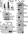
Fig. 1.
Fig. 1.
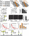
SKIP is an RI-selective AKAP. (A) HEK293 cells were transfected with vectors encoding FLAG-SKIP, FLAG-AKAP79, or FLAG-Trim63. FLAG immune complexes were analyzed for PKA activity in the presence or absence of PKI (means ± SEM, n = 4, **p ≤ 0.01, ***p ≤ 0.001). (B) FLAG immune complexes from HEK293 cells were immunoblotted with antibodies against the catalytic (Top), RI (Second), or RII (Third) subunits of PKA, or overlayed with purified labeled RI (Fourth) or RII (Fifth) protein. Immunoblot of input lysate is shown in Bottom. (C) Lysates from HEK293 cells expressing SKIP were immunoprecipitated with IgG, RI, or RII antibodies and immunoblotted for SKIP. Input lysate is shown in Bottom. (D) GST or GST-fused PKA regulatory subunits or fragments (indicated above each lane) were purified on glutathione sepharose. GST pull-downs were blotted for SKIP (Top) or AKAP79 (Bottom).
Fig. 2.
Fig. 2.
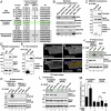
SKIP contains two independent RI-selective binding sites. (A) HEK293 cells were transfected with constructs for FLAG-AKAP79, FLAG-SKIP, or FLAG-SKIP lacking amino acids 900–950 (SKIP Δ1), amino acids 1,140–1,175 (SKIP Δ2), or amino acids 900–950 and 1,140–1,175 (SKIP Δ1,2). FLAG immune complexes were immunoblotted with antibodies against the RI (Top), RII (Second), or catalytic (Third) subunit of PKA. Input lysate is shown in lower three panels. (B) HEK293 cells were transfected with SKIP, SKIP Δ1, SKIP Δ2, SKIP Δ1,2, or Trim63. FLAG immune complexes were analyzed for PKA activity in the presence or absence of PKI (means ± SEM, n = 4, *p ≤ 0.01, ***p ≤ 0.001). (CD) Coomassie blue stain of affinity-purified FLAG-SKIP 750–1,100 (C) and FLAG-SKIP 1,100–1,400 (D). (EF) AlphaScreen assay performed with varying concentrations of purified FLAG-SKIP 750–1,100 (E) or FLAG-SKIP 1,100–1,400 (F) and biotinylated RI. Cross-titration was performed with anti-FLAG acceptor beads together with streptavidin donor beads. (GJ) Surface plasmon resonance analysis of FLAG-SKIP 750–1,100 (G) or FLAG-SKIP 1,100–1,400 (I) binding to immobilized RI. (H and J) Steady-state binding curves with dissociation constants (Kd) for interaction between SKIP 750–1,100 (H) or SKIP 1,100–1,400 (J) and RI. Data is representative of three independent experiments run on different surfaces (means ± SEM, n = 3).
Fig. 3.
Fig. 3.
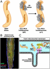
Stoichiometry and modeling of SKIP-RI interaction. (A) Alignment of amphipathic helices from three classes of AKAP protein. Residues are numbered according to the convention of previous structural studies (25, 26). (BC) Structural models of RI (gray). SKIP (orange) interactions are presented following superposition of SKIP residues 923–949 (B) and 1,145–1,171 (C) onto the equivalent region of D-AKAP2 in the D-AKAP2 PKA RI D/D crystal structure [Protein Data Bank (PDB) ID code 3IM4]. (DE) Alanine substitution of Phe929 and Tyr1151 abolishes RI binding in each GFP-fused binding fragment of SKIP (D) and significantly reduces RI binding in the context of full-length FLAG-SKIP (E). (F) Amalgamated densitometric analysis of RI binding to alanine substitution mutants of full-length FLAG-SKIP. (G) Representation of SiMPull assay performed to calculate stoichiometry of SKIP-RI complex. (HJ) YFP fluorescence from representative regions of different imaging surfaces. YFP fluorescence from anti-FLAG (H) or anti-GST (I) coated surface incubated with FLAG-SKIP and YFP-RI lysate or from surface alone not incubated with lysate (J). (K) Quantitation of YFP molecules on entire imaging surfaces from four independent experiments. (L) Representative decay time trajectories of a YFP spot requiring three or four photobleaching steps to quench YFP fluorescence. (M) Observed distribution of photobleaching steps from a representative experiment and predicted fit to two binomial distributions. (N) Schematic illustrating proportions of SKIP bound to one or two RI dimers as calculated by the SiMPull assay.
Fig. 4.
Fig. 4.
SKIP anchoring in RIα -/- mouse embryonic fibroblasts. (A) Wild-type MEFs transfected with FLAG-AKAP79 or FLAG-SKIP and FLAG immune complexes were immunoblotted with antibodies against RI (Top) or RII (Middle). Input lysate is shown in Bottom. (B) FLAG immune complexes were analyzed for PKA activity in the presence or absence of PKI (means ± SEM, n = 4, *p ≤ 0.05, **p ≤ 0.01). (C) RIα -/- MEFs expressing FLAG-SKIP-GFP or both FLAG-SKIP-GFP and V5-RI-mCherry were selected by flow cytometry. FLAG-SKIP immune complexes were immunoblotted with antibodies against RI (Top). Input lysate is shown in lower panels. (D) PKA activity measurements were performed in the presence or absence of PKI with FLAG immune complexes isolated from RIα -/- MEFs expressing FLAG-SKIP-GFP or FLAG-SKIP-GFP and V5-RI-mCherry (means ± SEM, n = 4, ***p ≤ 0.001).
Fig. 5.
Fig. 5.
SKIP and RI localize to mitochondria. (AD) COS-7 cells were transfected with GFP-SKIP (A, green), fixed, and then incubated with antibodies against RI (B, blue) or Mitofusin-1 (C, red). Composite image is shown in D. (EH) COS-7 cells expressing GFP-SKIP (E, green), were incubated with MitoTracker dye (250 nM) for 30 min prior to fixation (G, red), and then incubated with antibodies against RI (F, blue). Composite image is shown in H. (IL) Adult mouse cardiomyocytes were fixed and incubated with antibodies against SKIP (I, green), RI (J, blue), and Mitofusin-1 (K, red). Composite image is shown in L. Higher magnification images of adult mouse cardiomyocytes incubated with antibodies against SKIP (green, M, P), RI (red, N), or Mitofusin-1 (red, Q) are shown. Composite images are shown in O and R. (SU) Images of live GFP-SKIP expressing adult cardiomyocytes loaded with MitoTracker (red).
Fig. 6.
Fig. 6.
SKIP associates with and mediates PKA phosphorylation of ChChd3. (A) Mass spectrometry screening for mitochondrial SKIP-binding partners. FLAG-SKIP immune complexes isolated from HEK293 cells were proteolyzed and peptides were identified by MS/MS sequencing. Samples from SKIP immune complexes (SKIP) were compared to immune complexes from cells transfected with empty vector (empty). The name of each protein, the number of peptides identified, percent sequence coverage, and IP identification number are indicated above each column. Mitochondrial proteins are indicated. (B) Immunoblot detection of SKIP in mitochondrial subfractions. The name of each subfraction is indicated above each lane. Enrichment for the mitochondrial marker proteins VDAC (outer mitochondrial membranes), Smac (intermembrane space), NDUF A9 (inner mitochondrial membrane), and HSP70 (matrix) was used to assess quality of subfractionation. (C) HEK293 cells were transfected with V5-ChChd3 and either FLAG-AKAP79 or FLAG-SKIP. FLAG immune complexes were immunoblotted for V5-ChChd3 (Top). Input lysate is shown in Middle and Bottom. (D) V5-ChChd3 immune complexes were isolated and immunoblotted for FLAG-SKIP. Input lysate is shown in Middle and Bottom. (E) In vitro-translated V5-ChChd3 was incubated with in vitro-translated FLAG-SKIP or FLAG-AKAP79. FLAG immune complexes were immunoblotted for ChChd3 (Top). Input lysate is shown in Bottom. (FI) Immunofluorescence images of adult mouse cardiomyocytes incubated with antibodies against SKIP (F, green), ChChd3 (G, red), and RI (H, blue). The composite image is shown in I. (JM) Phosphorylation of ChChd3 by SKIP-anchored PKA. (J) HEK293 cells were transfected with FLAG-SKIP or FLAG-SKIP and V5-ChChd3. FLAG immune complexes were treated with vehicle or cAMP (100 μM) in the presence of γ-32P ATP. Phosphoproteins were resolved by SDS-PAGE and detected by autoradiography (Top). Input lysate is shown in Middle and Bottom. (K) HEK293 cells were cotransfected with V5-ChChd3 and either FLAG-SKIP or FLAG-SKIPΔ1,2. FLAG immune complexes were isolated and stimulated with vehicle, cAMP (100 μM), or cAMP and PKI, in the presence of γ-32P ATP. Phosphoproteins were resolved by SDS-PAGE and detected by autoradiography (Top). Input lysate is shown in Middle and Bottom. (L) HeLa cells were transfected with FLAG-SKIP and scrambled siRNA or siRNA against ChChd3. FLAG immune complexes were isolated and treated with vehicle, cAMP (100 μM), or cAMP and PKI in the presence of γ-32P ATP. Phosphoproteins were resolved by SDS-PAGE and detected by autoradiography (Top). Suppression of ChChd3 expression was assessed by immunoblot (Second). Input lysate and loading control are shown in Third and Bottom. (M) Amalgamated densitometric analysis of phosphate incorporation into ChChd3 in FLAG immune complexes isolated from HeLa cells expressing FLAG-SKIP (means ± SEM, n = 3, *p ≤ 0.05).
Fig. P1.
Fig. P1.
SKIP targets the type I PKA holoenzyme to mitochondria. (A) A model that depicts the RI anchoring sites on SKIP and the alternate stoichiometry of the SKIP∶PKA complexes. (B) The mitochondrial location of SKIP (green), RI (blue), and ChChd3 (red) in adult cardiomyocytes. (C) Cartoon depicting SKIP location and function in the intermembrane space of mitochondria.

References

    1. Keren H, Lev-Maor G, Ast G. Alternative splicing and evolution: Diversification, exon definition and function. Nat Rev Genet. 2010;11:345–355. - PubMed
    1. Hunter T. Protein kinases and phosphatases: The yin and yang of protein phosphorylation and signaling. Cell. 1995;80:225–236. - PubMed
    1. Manning G, Whyte DB, Martinez R, Hunter T, Sudarsanam S. The protein kinase complement of the human genome. Science. 2002;298:1912–1934. - PubMed
    1. Scott JD, Pawson T. Cell signaling in space and time: Where proteins come together and when they’re apart. Science. 2009;326:1220–1224. - PMC - PubMed
    1. Mitchison T, Kirschner M. Dynamic instability of microtubule growth. Nature. 1984;312:237–242. - PubMed

Publication types

MeSH terms

Substances