. 2011 Nov 29;108(48):19252-7.
doi: 10.1073/pnas.1116794108. Epub 2011 Nov 14.

Cell cycle adaptations of embryonic stem cells

Affiliations

Cell cycle adaptations of embryonic stem cells

Andrea Ballabeni et al. Proc Natl Acad Sci U S A. .

Abstract

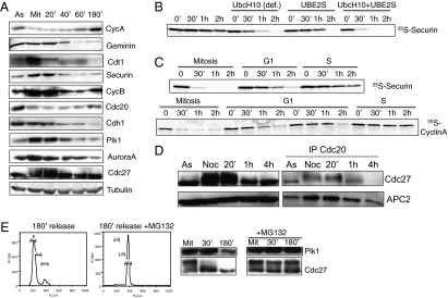
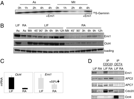
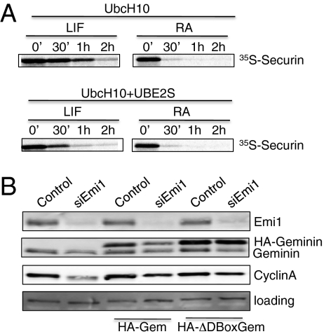
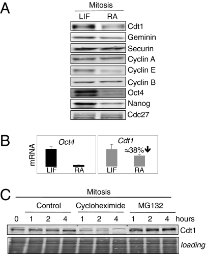
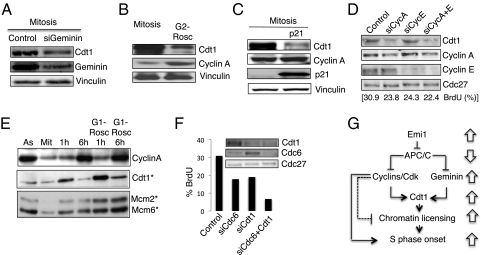
ES cells proliferate with very short gap phases yet maintain their capacity to differentiate. It had been thought that the levels of cyclins and other substrates of ubiquitin ligase APC/C remain nearly constant and Cdk activity remains constitutively high in mouse ES cells. Here we demonstrate that APC/C (anaphase-promoting complex/cyclosome) enzyme is active in ES cells but attenuated by high levels of the Emi1 (early mitotic inhibitor-1) protein. Despite the presence of high Cdk activity during the G1 phase, chromatin can be effectively licensed for DNA replication and fast entry into the S phase can still occur. High Cdk activity during S-G2-M phases produces high levels of the DNA replication factor Cdt1, and this leads to efficient Mcm proteins loading on chromatin after mitotic exit. Although disturbing the usual balance between Cdk activity and APC/C activity found in somatic cells, a few key adaptations allow normal progression of a very rapid cell cycle.


Conflict of interest statement

The authors declare no conflict of interest.

Figures

Fig. 1.
Fig. 1.
APC/C activity oscillates in mouse embryonic stem cells. (A) Immunoblotting analysis for the indicated proteins from cells synchronized as shown in SI Appendix, Fig. S2. Cdt1 is partially degraded and migrating faster in the G1 phase; this pattern recapitulates somatic cells (13, 27) and the slower migration in SDS gel during mitosis is plausibly because of phosphorylation. In addition, Cdc27 and Cdh1 are phosphorylated during mitosis. (B) Degradation assay from mitotic ES cell extracts. Degradation assay is performed at 30 °C for the indicated times and in vitro-translated 35S-Securin is used as a substrate. E2 enzymes UbcH10 and UBE2S are purified as previously shown (5). Autoradiography for 35S-Securin is shown. (C) Degradation assays with extracts from cells synchronized at different phases of the cell cycle. (Upper) Degradation assay from CCE ES cells synchronized at mitosis, 1 h, and 4 h, released from mitosis with in vitro-translated 35S-Securin as a substrate. (Lower) Degradation assay from J1 ES cells synchronized at mitosis, 1 h, 1 h 30 min, and 4 h, released from mitosis with in vitro-translated 35S-Cyclin A as a substrate. Autoradiographies for 35S-Securin or 35S-Cyclin A are shown. (D) Cdc20 dissociates from the APC/C complex after mitotic exit. ES cells synchronized in mitosis and released for the indicated times are used to perform immunoprecipitation of Cdc20. Immunoblotting analysis for the indicated coimmunoprecipitated proteins is shown. (E) Proteasome inhibition blocks exit from mitosis and APC/C-dependent degradation. ES cells are released in presence or absence of proteasome inhibitor MG132 for 3 h. FACS analyses (Left) and immunoblotting (Right) for APC/C subunit Cdc27 and APC/C substrate Plk1.
Fig. 2.
Fig. 2.
Emi1 levels are high in ES cells. (A) Emi1 inhibits APC/C activity in ES cells. Degradation assay is performed with nonsynchronized or mitotic ES cells for the indicated times. In vitro translated 35S-Geminin is used as a substrate. Recombinant N-terminal–deleted Emi1 is used to inhibit the degradation. Autoradiography for 35S-Securin is shown. (B) Emi1 protein levels are decreased upon differentiation. ES cells are maintained in leukemia inhibitory factor (LIF) containing medium or treated with RA for 48 h to induce differentiation. Cells are synchronized in mitosis and released for the indicated times. Immunoblotting analysis for the indicated proteins is shown. Oct4 is used as a marker for pluripotency. (C) RNA levels for Emi1 are decreased during differentiation. RNA levels for Emi1 and Oct4 are determined by quantitative PCR (qPCR) performed on three independent biological samples synchronized at different time points during S-G2-M progression. Reduction in mRNA for Emi1 is shown. Samples are normalized to β-actin. (D) Emi1 levels associated to APC/C are decreased in differentiated cells. Control cells and cells treated with RA are synchronized in mitosis and immunoprecipitations of Cdc20 or Oct4 (negative control) are performed before immunoblotting analysis for the indicated proteins. Lanes 1 and 2 correspond to 2.5% input.
Fig. 3.
Fig. 3.
APC/C activity is increased upon differentiation. (A) Degradation assay from undifferentiated ES cells or ES cells induced to differentiation for 48 h with RA is shown. In vitro translated 35S-Securin is used as a substrate. Recombinant UbcH10 alone or UbcH10 in combination with UBE2S were used for the comparisons. These data are from same experiment as shown in Fig. 1B. Autoradiography for 35S-Secruin is shown. (B) Emi1 depletion leads to decrease in APC/C substrates levels. Control or siRNA for Emi1 are transfected in ES cells using Lipofectamine 2000 transfection reagent (Invitrogen). Thirty hours after transfection, levels of the indicated proteins are evaluated by immunoblotting analysis. In lanes 3–6, the indicated HA-tagged versions of Geminin are transfected 6 h after siRNA transfection using FuGene 6 reagent (Promega).
Fig. 4.
Fig. 4.
Cdt1 is abundant in ES cells. (A) Cdt1 protein levels are higher in mitotic ES cells than in differentiated cells. Immunoblotting for the indicated proteins is shown. Oct4 and Nanog are used as markers of pluripotency. Cdc27 phosphorylation is used as a marker of equal synchronization in mitosis. Mitotic extract of ES cells and differentiated cells (after standard 48-h treatment with RA) are used. (B) RNA levels for Cdt1 are decreased during differentiation. RNA levels for Cdt1 and Oct4 are determined by qPCR performed on three independent biological samples synchronized at different time points during S-G2-M progression. Reduction in mRNA level for Cdt1 is shown. Samples are normalized to β-actin. (C) Cdt1 turnover in mitosis is fast and protein levels are maintained high. Mitotic ES cells are treated with protein-synthesis inhibitor cycloheximide or proteasome inhibitor MG132 for the indicated times. Immunoblotting for Cdt1 protein is shown and equal loading is evaluated by Ponceau staining.
Fig. 5.
Fig. 5.
Geminin and Cdk activity amplify Cdt1 protein levels. (A) siRNA for Geminin is transfected 1 h after release from thymidine arrest and in the presence of Nocodazole for synchronization in mitosis 7 h later. Immunoblotting analysis for the indicated proteins is shown. Vinculin is used as a loading control. (B) Roscovitine treatment in the G2 phase blocks cell-cycle progression at G2/M transition with lower levels of Cdt1 protein. ES cells released for 1 h after thymidine block are treated with Cdk1/Cdk2 inhibitor Roscovitine or DMSO (control) and incubated for other 6 h in the presence of Nocodazole. Immunoblotting for the indicated proteins is shown. Vinculin is used as a marker of equal loading. (C) Cdt1 is decreased in mitosis when Cdk inhibitor p21 is expressed. Cells expressing p21 by Doxycycline treatment are used together with control cells (i.e. not treated with Doxycycline) and synchronized in mitosis. Immunoblotting analysis for the indicated proteins is shown. (D) Cyclin A depletion reduces levels of Cdt1. ES cells released for 1 h from thymidine arrest are treated with the indicated siRNA for 7 h in the presence of Nocodazole. The cells are then released from Nocodazole block and immunoblotting for the indicated proteins is performed with lysates from cells released for 1 h. BrdU incorporation was evaluated 2 h 45 min after Nocodazole release (15-min labeling). (E) Cdk activity during the G1 phase inhibits pre-RC formation. ES cells are synchronized in mitosis before release and treatment with Roscovitine during G1 phase. Immunoblotting analysis for the level of indicated proteins is shown. The asterisk represents the chromatin-containing fraction. (F) Cdt1 and Cdc6 drive fast G1 progression in ES cells. ES cells are synchronized in the S phase by thymidine treatment and released in presence of Nocodazole and siRNA for Cdt1 and Cdc6. Cells are then released from mitosis for 2 h 45 min with the last 15 min labeling with BrdU. Quantification of BrdU-positive cells is shown. (Inset) Immunoblotting analysis for levels of the indicated proteins in the siCdc6 or/and siCdt1 treated cells. (G) Model for the regulation of fast S phase entry in ES cells. Block arrows represent comparison with somatic cells.

References

    1. White J, et al. Developmental activation of the Rb-E2F pathway and establishment of cell cycle-regulated cyclin-dependent kinase activity during embryonic stem cell differentiation. Mol Biol Cell. 2005;16:2018–2027. - PMC - PubMed
    1. Savatier P, Lapillonne H, Jirmanova L, Vitelli L, Samarut J. Analysis of the cell cycle in mouse embryonic stem cells. Methods Mol Biol. 2002;185:27–33. - PubMed
    1. Fujii-Yamamoto H, Kim JM, Arai K, Masai H. Cell cycle and developmental regulations of replication factors in mouse embryonic stem cells. J Biol Chem. 2005;280:12976–12987. - PubMed
    1. Bloom J, Cross FR. Multiple levels of cyclin specificity in cell-cycle control. Nat Rev Mol Cell Biol. 2007;8:149–160. - PubMed
    1. Wu T, et al. UBE2S drives elongation of K11-linked ubiquitin chains by the anaphase-promoting complex. Proc Natl Acad Sci USA. 2010;107:1355–1360. - PMC - PubMed

Publication types

LinkOut - more resources