. 2011 Dec 6;108(49):E1330-8.
doi: 10.1073/pnas.1110013108. Epub 2011 Nov 14.

Diameter and rigidity of multiwalled carbon nanotubes are critical factors in mesothelial injury and carcinogenesis

Affiliations

Diameter and rigidity of multiwalled carbon nanotubes are critical factors in mesothelial injury and carcinogenesis

Hirotaka Nagai et al. Proc Natl Acad Sci U S A. .

Abstract

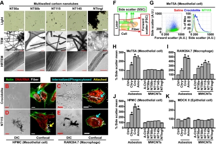
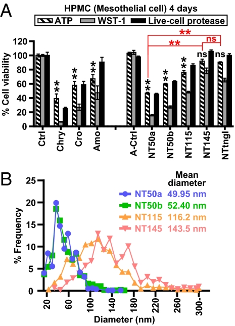
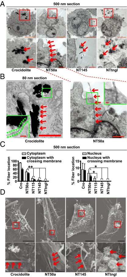
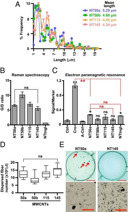
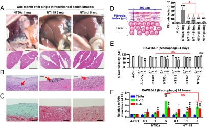
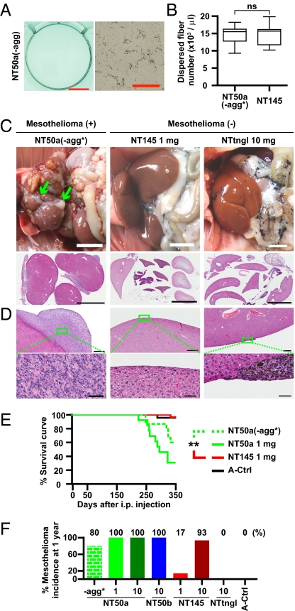
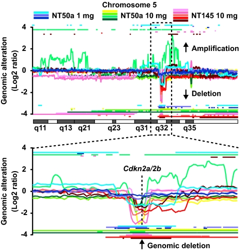
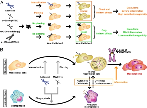
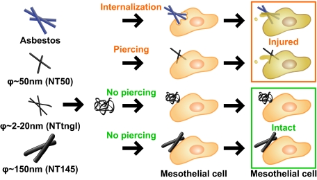
Multiwalled carbon nanotubes (MWCNTs) have the potential for widespread applications in engineering and materials science. However, because of their needle-like shape and high durability, concerns have been raised that MWCNTs may induce asbestos-like pathogenicity. Although recent studies have demonstrated that MWCNTs induce various types of reactivities, the physicochemical features of MWCNTs that determine their cytotoxicity and carcinogenicity in mesothelial cells remain unclear. Here, we showed that the deleterious effects of nonfunctionalized MWCNTs on human mesothelial cells were associated with their diameter-dependent piercing of the cell membrane. Thin MWCNTs (diameter ∼ 50 nm) with high crystallinity showed mesothelial cell membrane piercing and cytotoxicity in vitro and subsequent inflammogenicity and mesotheliomagenicity in vivo. In contrast, thick (diameter ∼ 150 nm) or tangled (diameter ∼ 2-20 nm) MWCNTs were less toxic, inflammogenic, and carcinogenic. Thin and thick MWCNTs similarly affected macrophages. Mesotheliomas induced by MWCNTs shared homozygous deletion of Cdkn2a/2b tumor suppressor genes, similar to mesotheliomas induced by asbestos. Thus, we propose that different degrees of direct mesothelial injury by thin and thick MWCNTs are responsible for the extent of inflammogenicity and carcinogenicity. This work suggests that control of the diameter of MWCNTs could reduce the potential hazard to human health.


Conflict of interest statement

The authors declare no conflict of interest.

Figures

Fig. 1.
Fig. 1.
Mesothelial cells did not phagocytose MWCNTs. (A) Optical and transmission EM (TEM) images and high-resolution (HR) Transmission EM images for five types of MWCNTs. Each carbon nanotube is named after its diameter (Fig. 2 and Table S1) except NTtngl, which stands for tangled NTs. High-resolution transmission EM images show the presence of graphite layers in the MWCNTs. (Scale bars: light microscopy, 20 μm; all transmission EM images except NTtngl, 500 nm; NTtngl transmission EM image, 200 nm; and high-resolution transmission EM images, 10 nm.) (B and D) Differential interference contrast and confocal Images of HPMCs show Cro internalization (blue arrow) and NT115 attachment (yellow arrow) after a 24-h incubation. (C and E) Images of RAW264.7 cells show internalization of both fibers (blue arrows). (BE) Differential interference contrast images (Left) and confocal microscopy images (Right) after staining of the cells with Alexa 488-phalloidin (green) and propidium iodide (PI, red), indicate cytoskeletal actin and DNA/RNA, respectively. (Scale bars: BE, 14 μm.) (F) A schematic showing the SSC and forward scatter (FSC) values for uptake of fibers. (G) Left: Dot plot of forward scatter and SSC values of MeT5A cells show a marked increase in SSC only upon incubation with Cro, but not with NT115. Right: Histogram shows SSC value of the image on the left. (HK) Mean of the SSC value of each cell after a 3-h incubation with each fiber type (*P < 0.001; n = 3, mean ± SEM). P values were calculated between control (saline) and asbestos, or A-control (A-saline solution) and MWCNTs.
Fig. 2.
Fig. 2.
Diameter of MWCNTs inversely correlated with mesothelial toxicity. (A) Cell viability assays of HPMCs after a 4-d incubation with each fiber at 5 μg/cm2 show variable mesothelial cell cytotoxicity by MWCNTs. NT50a showed strong cytotoxicity against mesothelial cells, whereas NT145 and NTtngl did not. Ctrl, control. P values were calculated between control (saline) and asbestos or A-control (A-saline solution) and MWCNTs (n = 3, mean ± SEM; **P < 0.01; ns, not significant). (B) Distribution of the diameters of MWCNTs.
Fig. 3.
Fig. 3.
Thin, dispersed MWCNTs with high crystallinity pierced mesothelial cells. (A) Representative transmission EM images of HPMCs after a 3-h incubation with indicated fibers. Ultrathin sections were prepared at a thickness of 500 nm. Squares (Upper) indicate insets (Lower). Cro was fully internalized. NT50a penetrated the mesothelial cell membrane, but NT145 and NTtngl did not (red arrow). (Scale bars: Upper, 10 μm; Lower, 1 μm.) (B) Cro was internalized and surrounded by a vesicular membrane structure (Left), whereas NT50a pierced the plasma membrane of the mesothelial cell without a vesicular structure surrounding the nanotubes (Right). We prepared 80-nm sections to show vesicular structure. Green arrowhead indicates vesicular structure. (Scale bars: 1 μm; Insets, 100 nm.) (C) Localization of fibers in transmission EM images (n = 3, mean ± SEM; *P < 0.05 and **P < 0.01). P values were calculated by Student's t test. (D) Representative scanning EM images of HPMCs after a 3-h incubation with indicated fibers.
Fig. 4.
Fig. 4.
Other factors, including length, structural defects, free radical generation, and difference in number of fibers, were not the primary causes of mesothelial cell cytotoxicity by thin, dispersed MWCNTs with high crystallinity. (A) Distribution of the lengths of MWCNTs. (B) Graphite/defect (G/D) ratio shows no significant difference in defect abundance between NT50a and NT145. (C) EPR shows that NT50a and NTtngl produce hydroxyl radicals in the presence of hydrogen peroxide, whereas NT145 does not. Cro was most potent in free radical generation. P values were calculated between control (saline) and asbestos or A-control (A-saline) and MWCNTs. (D) Number of dispersed fibers in 0.5 mg/mL fiber suspensions. (E) Macroscopic and microscopic images of NT50a and NT145 suspensions under experimental conditions (5 μg/cm2). Arrows indicate nanotube aggregation, which greatly reduces the suspension of dispersed fibers. (Scale bars: Upper, 10 mm; Lower, 100 μm.) P values (*P < 0.05 and **P < 0.01; ns, not significant) were determined by one-way ANOVA with post-hoc Dunnett test (C) or Tukey test (B and D).
Fig. 5.
Fig. 5.
Thin, dispersed MWCNTs with high crystallinity caused severe fibrotic peritonitis in rats, although affected macrophages in vitro similarly compared with the other MWCNTs. (AC) Macroscopic and microscopic images of rat organs 1 mo after a single i.p. injection of 1 mg NT50a (Left), 5 mg NT145 (Middle), or 5 mg NTtngl (Right). (A) Macroscopic images of i.p. organs immediately after dissection (Top). Low-power view of H&E-stained livers show an altered dull edge (Lower Left). (Scale bar: 1 cm.) (B) Microscopic images of the liver surface show the deposition of fibers (arrow) and various fibrotic responses around fibers. (Scale bars: 50 μm.) (C) Masson trichrome stain showing abundant light green collagen (Left). (Scale bars: 50 μm.) (D) Fibrosis index was calculated by measuring the distance from the entire outer surface of the liver to the mesothelium at intervals of 500 μm (Left). Fibrosis index shows that NT50a induced the strongest fibrotic inflammation [Right; n = 3–6, mean ± SEM; *P < 0.05; ns, not significant between A-control (A-saline) and MWCNT]. (E) Cell viability assays of macrophages (RAW264.7) after a 4-d incubation with each fiber at indicated concentrations (0.1, 1, or 5 μg/cm2) show that there was no significant difference in cytotoxic effects between NT50a and NT145. (F) Macrophages showed increased mRNA levels of IL-1β and IL-6 after a 24-h incubation with NT50a and NT145 at 5 μg/cm2, and there was no significant difference between NT50a and NT145 for mRNA of inflammatory cytokines (n = 3–6, mean ± SEM; *P < 0.05 and **P < 0.01; ns, not significant). P values were determined by one-way ANOVA with post-hoc Tukey test (D and E) or by Student's t test (F).
Fig. 6.
Fig. 6.
Thin, dispersed MWCNTs with high crystallinity had higher mesotheliomagenicity than thick MWCNTs when injected intraperitoneally into rats. (A) Macroscopic and microscopic images of NT50a(-agg). NT50a(-agg) consisted of the supernatant of the original NT50a suspension after centrifugation. No agglomeration can be seen (compare vs. Fig. 4E). (Scale bars: Left, 10 mm; Right, 100 μm.) (B) NT50a(-agg*) had twice as many fibers in suspension as NT50a(-agg). The number of dispersed fibers in NT50a(-agg*) and NT145 was almost the same (ns, not significant). The text provides further details. (C) Macroscopic images of i.p. organs immediately after dissection (Upper). Mesothelioma covers liver surface (Upper Left). Low-power view of H&E-stained livers shows malignant mesothelioma (Lower Left) or absence of mesothelioma (Lower Middle, Lower Right). (Scale bars: 1 cm.) (D) Microscopic images of the liver surface show mesothelioma (Left). Granuloma is seen (Right), and squares (Upper) indicate insets (Lower). (Scale bars: Upper, 500 μm; Lower, 50 μm.) (E) Survival curve of rats injected with MWCNTs and vehicle control shows that there was significant difference in mesotheliomagenicity between NT50a(-agg*) and NT145 (**P < 0.01). (F) Overall tumor probability induced by each fiber during the 1-y observation period. The number below the x axis indicates the amount of injected fibers [in mg; n = 15 in NT50a(-agg*), n = 13 in 1 mg NT50a, n = 43 in 10 mg NT50a, n = 6 in 10 mg NT50b, n = 29 in 1 mg NT145, n = 30 in 10 mg NT145, n = 15 in 10 mg NTtngl, and n = 23 in A-saline solution (A-Ctrl)]. Table S2 includes the numbers of rats used and histology of mesothelioma. P values were determined by Student’s t test (B) or by log-rank test (E).
Fig. 7.
Fig. 7.
Cdkn2a/2b was homozygously deleted in MWCNT-induced mesothelioma of rats. Array-based CGH analysis of mesotheliomas shows that the genomic locus, Cdkn2a/2b, was homozygously or heterozygously deleted in all mesothelioma cases tested. Horizontal lines above and below the CGH ratio curve were delineated based on z-score (Materials and Methods), indicating amplification and deletion of the indicated loci, respectively. At the Cdkn2a/2b locus, all tumors have horizontal lines below the CGH curve and thus have genomic deletion. Fig. S6 shows signal intensity and extent of genomic deletion at the Cdkn2a/2b locus.
Fig. 8.
Fig. 8.
Schematic of the effect of different MWCNTs on mesothelial cells. (A) The present study shows that asbestos and thin, dispersed MWCNTs with high crystallinity (diameter∼ 50 nm; .e.g., NT50a) penetrate mesothelial cells and further induce cell injury. However, aggregative MWCNTs (diameter ∼ 2–20 nm; e.g., NTtngl) and thick MWCNTs (diameter ∼ 150 nm; e.g., NT145) do not penetrate mesothelial cells, and thus there is no cell injury, which may explain the difference in the carcinogenicity between thin and thick MWCNTs. (B) Current schematic for mechanisms of mesotheliomagenesis focused on mesothelial injury and macrophage activation. Interplay of these two factors may lead to persistent inflammation and subsequent mesotheliomagenesis.
Fig. P1.
Fig. P1.
Effects of MWCNTs of different diameters on mesothelial cells. The present study shows that thin MWCNTs with high rigidity (e.g., NT50a) penetrate mesothelial cells and further induce cell injury. However, thick MWCNTs (e.g., NT145) do not penetrate mesothelial cells, and thus cause no cell injury. The mesothelial toxicity induced by thin and rigid MWCNT types may contribute to inflammation and carcinogenesis.

References

    1. Iijima S. Helical microtubules of graphitic carbon. Nature. 1991;354:56–58.
    1. Thayer AM. Carbon nanotubes by the metric ton. Chem Eng News. 2007;85:29.
    1. Sakamoto Y, et al. Induction of mesothelioma by a single intrascrotal administration of multi-wall carbon nanotube in intact male Fischer 344 rats. J Toxicol Sci. 2009;34:65–76. - PubMed
    1. Takagi A, et al. Induction of mesothelioma in p53+/- mouse by intraperitoneal application of multi-wall carbon nanotube. J Toxicol Sci. 2008;33:105–116. - PubMed
    1. Donaldson K, Poland CA. Nanotoxicology: New insights into nanotubes. Nat Nanotechnol. 2009;4:708–710. - PubMed

Publication types

MeSH terms

Substances