. 2011 Nov 29;108(48):19341-6.
doi: 10.1073/pnas.1110946108. Epub 2011 Nov 14.

Tripartite motif 8 (TRIM8) modulates TNFα- and IL-1β-triggered NF-κB activation by targeting TAK1 for K63-linked polyubiquitination

Affiliations

Tripartite motif 8 (TRIM8) modulates TNFα- and IL-1β-triggered NF-κB activation by targeting TAK1 for K63-linked polyubiquitination

Qi Li et al. Proc Natl Acad Sci U S A. .

Abstract

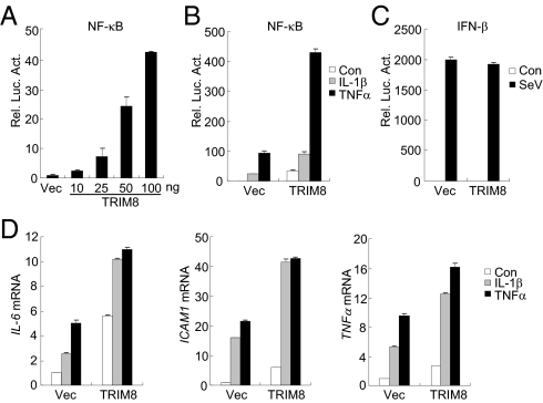
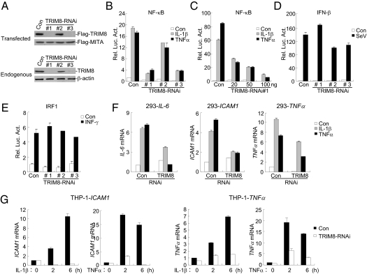
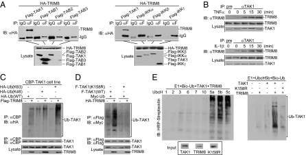
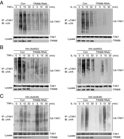
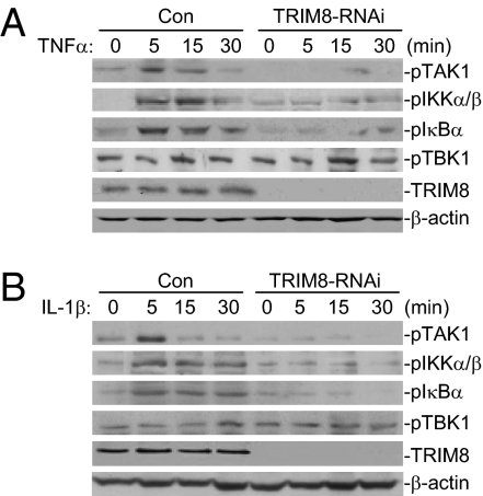
The tripartite motif (TRIM)-containing proteins are a family of proteins that have been known to be involved in divergent biological processes, including important roles in immune responses through regulating various signaling pathways. In this study, we identified a member of the TRIM family, TRIM8, as a positive regulator of tumor necrosis factor-α (TNFα) and interleukin-1β (IL-1β)-triggered NF-κB activation. Overexpression of TRIM8 activated NF-κB and potentiated TNFα- and IL-1β-induced activation of NF-κB, whereas knockdown of TRIM8 had opposite effects. Coimmunoprecipitations indicated that TRIM8 interacted with TGFβ activated kinase 1 (TAK1), a serine/threonine kinase essential for TNFα- and IL-β-induced NF-κB activation. Furthermore, we found that TRIM8 mediated K63-linked polyubiquitination of TAK1 triggered by TNFα and IL-1β. Our findings demonstrate that TRIM8 serves as a critical regulator of TNFα- and IL-1β-induced NF-κB activation by mediating K63-linked polyubiquitination of TAK1.


Conflict of interest statement

The authors declare no conflict of interest.

Figures

Fig. 1.
Fig. 1.
Identification of TRIM8 as a positive regulator of TNFα- and IL-1β–triggered NF-κB and transcription induction of inflammatory genes. (A) Overexpression of TRIM8 activates NF-κB in a dose-dependent manner. A total of 293 cells (1 × 10⁵) were transfected with NF-κB reporter (0.01 μg) and an increased amount of TRIM8 expression plasmid. Luciferase assays were performed 20 h after transfection. (B) Effects of TRIM8 on TNFα- and IL-1β–induced activation of NF-κB. A total of 293 cells (1 × 10⁵) were transfected with NF-κB reporter (0.01 μg) and empty vector or TRIM9 expression plasmid. Twenty hours after transfection, cells were left untreated or stimulated with TNFα (10 ng/mL) or IL-1β (10 ng/mL) for 10 h before luciferase assays were performed. (C) TRIM8 is not involved in SeV-induced activation of the IFNβ promoter. Reporter assays were performed as described in B, except that the IFNβ promoter reporter plasmid was used. (D) TRIM8 potentiates TNFα- and IL-1β–induced transcription of endogenous IL-6, ICAM1, and TNFα genes. A total of 293 cells (2 × 10⁵) were transfected with an empty vector or TRIM8 expression plasmid (0.1 μg). Twenty hours after transfection, cells were left untreated or stimulated with TNFα (10 ng/mL) or IL-1β (10 ng/mL) for 10 h before quantitative RT-PCR was performed.
Fig. 2.
Fig. 2.
Knockdown of TRIM8 inhibits TNFα- and IL-1β–induced NF-κB activation. (A) Effects of TRIM8 RNAi plasmids on expression of TRIM8. (Upper) A total of 293 cells (2 × 10⁵) were transfected with Flag-TRIM8 (0.2 μg each), Flag-MITA (0.05 μg), and the indicated RNAi (1 μg) plasmids. Thirty-six hours later, immunoblot analysis was performed with anti-Flag. (Lower) A total of 293 cells (2 × 10⁵) were transfected with the indicated RNAi plasmids (1 μg) for 48 h, and then immunoblot analysis was performed with the indicated antibodies. (B) Effects of TRIM8 RNAi plasmids on TNFα- and IL-1β–induced NF-κB activation. A total of 293 cells (1 × 10⁵) were transfected with NF-κB reporter (0.01 μg) and the indicated RNAi plasmids. Thirty-six hours after transfection, cells were left untreated or stimulated with TNFα (10 ng/mL) or IL-1β (10 ng/mL) for 10 h before luciferase assays were performed. (C) TRIM8-RNAi inhibits TNFα- and IL-1β–induced activation of the NF-κB in a dose-dependent manner. A total of 293 cells (1 × 10⁵) were transfected with NF-κB reporter (0.01 μg) and an increased amount of the #1 TRIM8-RNAi plasmid. Reporter assays were performed as described in B. (D and E) Effects of TRIM8 RNAi on SeV-triggered activation of the IFNβ promoter (D) or IFNγ-induced activation of the IRF1 promoter (E). A total of 293 cells (1 × 10⁵) were transfected with the indicated reporter (0.05 μg) and TRIM-RNAi plasmids (0.5 μg) for 36 h. Cells were then subjected to the indicated stimulation for 8 h before luciferase assays were performed. (F) Effects of TRIM8 RNAi on TNFα- or IL-1β–induced transcription of endogenous IL-6, ICAM1, and TNFα genes in 293 cells. These 293 cells (2 × 10⁵) were transfected with the indicated RNAi plasmids (2 μg). Thirty-six hours after transfection, cells were left untreated or stimulated with TNFα (10 ng/mL) or IL-1β (10 ng/mL) for 10 h before RT-PCR experiments were performed. (G) Effects of TRIM8 RNAi on TNFα- or IL-1β–induced transcription of endogenous ICAM1 and TNFα genes in THP-1 cells. THP-1 cells were transduced with GFP control or TRIM8 RNAi by retrovirus-mediated gene transfer. The cells were then either untreated or treated with TNFα (10 ng/mL) or IL-1β (10 ng/mL) for the indicated times before RT-PCR experiments were performed.
Fig. 3.
Fig. 3.
TRIM8 interacts with and ubiquitinates TAK1. (A) TRIM8 interacts with TAK1 in the mammalian overexpression system. A total of 293 cells (2 × 10⁶) were transfected with the indicated plasmids (5 μg each), and coimmunoprecipitation and immunoblot analysis were performed with the indicated antibodies (Upper panels). Expression of the transfected proteins was analyzed by immunoblots with anti-HA and anti-Flag (Lower panels). (B) Endogenous TRIM8 is associated with TAK1 following TNFα or IL-1β stimulation. A total of 293 cells (2 × 10⁷) were left untreated or stimulated with TNFα or IL-1β for the indicated times. Immunoprecipitation and immunoblot analysis were performed with the indicated antibodies. (C) Overexpressed TRIM8 enhances wild-type and K63-linked polyubiquitination of TAK1. A total of 293 cells (2 × 10⁶) stably expressing calmodulin-binding peptide (CBP)-tagged TAK1 were transfected with Flag-TRIM8 and the indicated ubiquitin plasmids. Twenty-four hours after transfection, cell lysates were immunoprecipitated with anti-CBP. The immunoprecipitates were analyzed by immunoblots with anti-HA, anti-TAK1, or anti-Flag as indicated. (D) TRIM8 enhances polyubiquitination of wild-type TAK1 but not of TAK1(K158R) in vivo. A total of 293 cells (2 × 10⁶) were transfected with the indicated plasmids. Twenty-four hours after transfection, cell lysates were immunoprecipitated with anti-Myc. The immunoprecipitates were analyzed by immunoblots with anti-HA or anti-Flag as indicated. (E) TRIM8 enhances polyubiquitination of wild-type TAK1 but not of TAK1(K158R) in vitro. TAK1, TAK1(K158R), and TRIM8 were translated in vitro, and biotin–ubiquitin, E1, and the indicated E2s were added for ubiquitination assays. Ubiquitin-conjugated proteins were detected by immunoblot with HRP–streptavidin. Before ubiquitination analysis, the input levels of the translated proteins were detected by immunoblots.
Fig. 4.
Fig. 4.
TRIM8-RNAi reduces TNFα- and IL-1β–induced K63-linked polyubiquitination of endogenous TAK1. (A) Effects of TRIM8 RNAi on TNFα- or IL-1β–induced polyubiquitination of endogenous TAK1. A total of 293 cells stably transduced with a control or TRIM8 RNAi were either untreated or stimulated with TNFα (10 ng/mL) or IL-1β (10 ng/mL) for the indicated times. Cell lysates were immunoprecipitated with anti-TAK1, and the immunoprecipitates were analyzed by immunoblots with anti-ubiquitin, anti-TRIM8, or anti-TAK1 as indicated. (B and C) Effects of TRIM8 RNAi on TNFα- or IL-1β–induced K63-linked (B) or K48-linked (C) polyubiquitination of endogenous TAK1. A total of 293 cells stably transduced with a control or TRIM8 RNAi were transfected with HA-tagged K63-only or K48-only ubiquitin plasmid. Twenty-four hours after transfection, cell lysates were immunoprecipitated with anti-TAK1. The immunoprecipitates were analyzed by immunoblots with anti-HA, anti-TAK1, and anti-TRIM8 as indicated.
Fig. 5.
Fig. 5.
Knockdown of TRIM8 reduces TNFα- and IL-1β–induced phosphorylation of TAK1, IKKα/β, and IκBα. (A and B) TRIM8-RNAi inhibits TNFα-induced phosphorylation of TAK1, IKKα/β, IKBα but not of TBK1. A total of 293 cells stably transduced with a control or TRIM8 RNAi plasmid were either untreated or treated with TNFα (10 ng/mL) (A) or IL-1β (10 ng/mL) (B) for the indicated times and then subsequently analyzed by immunoblots with the indicated antibodies.

References

    1. Hayden MS, Ghosh S. NF-κB in immunobiology. Cell Res. 2011;21:223–244. - PMC - PubMed
    1. Vallabhapurapu S, Karin M. Regulation and function of NF-kappaB transcription factors in the immune system. Annu Rev Immunol. 2009;27:693–733. - PubMed
    1. Chen ZJ. Ubiquitin signalling in the NF-kappaB pathway. Nat Cell Biol. 2005;7:758–765. - PMC - PubMed
    1. Jiang Z, Ninomiya-Tsuji J, Qian Y, Matsumoto K, Li X. Interleukin-1 (IL-1) receptor-associated kinase-dependent IL-1-induced signaling complexes phosphorylate TAK1 and TAB2 at the plasma membrane and activate TAK1 in the cytosol. Mol Cell Biol. 2002;22:7158–7167. - PMC - PubMed
    1. Yamaguchi K, et al. Identification of a member of the MAPKKK family as a potential mediator of TGF-beta signal transduction. Science. 1995;270:2008–2011. - PubMed

Publication types

MeSH terms

LinkOut - more resources