. 2011 Dec 13;108(50):20008-11.
doi: 10.1073/pnas.1116965108. Epub 2011 Nov 14.

Acrosome-reacted mouse spermatozoa recovered from the perivitelline space can fertilize other eggs

Affiliations

Acrosome-reacted mouse spermatozoa recovered from the perivitelline space can fertilize other eggs

Naokazu Inoue et al. Proc Natl Acad Sci U S A. .

Abstract

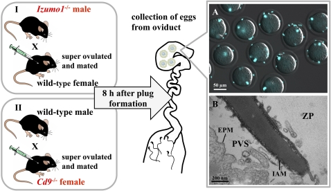
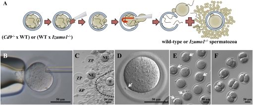
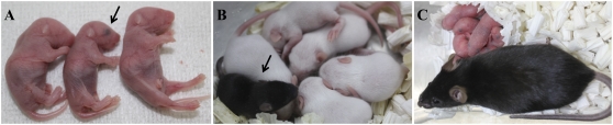
Many investigators maintain that spermatozoa that have initiated the acrosome reaction (AR) before reaching the surface of the egg's zona pellucida (ZP) are unable to bind and penetrate the ZP. A recent study has revealed that most fertilizing mouse spermatozoa initiate the AR before contacting the ZP. We found that acrosome-reacted spermatozoa collected from the perivitelline space of Cd9-null mice (whose egg plasma membranes are incapable of fusing with spermatozoa) were able to pass through both the cumulus and ZP of WT mouse eggs and produced live offspring. This means that the spermatozoa we used had the ability to pass through the ZP at least twice. Apparently, some spermatozoa that had undergone the AR long before contact with the ZP remained capable of crossing the ZP and fertilizing eggs. Thus, the concept that acrosome-reacted spermatozoa are unable to bind to the ZP and have lost their fertilizing capacity must be reconsidered.


Conflict of interest statement

The authors declare no conflict of interest.

Figures

Fig. 1.
Fig. 1.
Preparation of spermatozoa accumulating in the PVS. Eggs from either B6D2F1 or Cd9−/− female mice were superovulated as indicated in Materials and Methods. One hour before the estimated ovulation time, both lines of female mice were mated with Izumo1−/− or B6D2F1 male mice, respectively. Eggs were collected from oviducts 8 h after plug formation. (A) Many spermatozoa accumulated in the PVS of Cd9−/− eggs because of impaired sperm–egg fusion. The spermatozoa were stained with Hoechst 33342 and viewed through a fluorescence microscope with phase-contrast optics. A mean of 5.5 spermatozoa per egg was able to penetrate the zona (n = 11). (Scale bar = 50 μm.) (B) Transmission electron micrograph of a spermatozoon within the PVS; the micrograph shows a longitudinal section of the anterior region of the sperm head. The inner acrosomal membrane (IAM) is exposed. Vesicle-like structures around the IAM are sections of egg's microvilli. EPM, egg-plasma membrane. (Scale bar = 200 nm.)
Fig. 2.
Fig. 2.
Recovery of spermatozoa from the PVS and subsequent fertilization in vitro. (A) Schematic diagram indicating the recovery of spermatozoa from the PVS and subsequent in vitro insemination and fertilization. (B) Cumulus cells and all the spermatozoa near and on the ZP were removed by hyaluronidase treatment and subsequent washings. A few spermatozoa on the ZP, if any, were individually immobilized (permanently) by damaging sperm tail plasma membrane by a single piezo pulse. A large slit was made in the ZP by applying several piezo pulses to the pipette before spermatozoa in the PVS were flushed out from the slit together with the ooplasm (Movie S1). (C) Second round of insemination was performed in a 4-μL drop of TYH medium. The spermatozoa released from the PVS were coincubated with a freshly prepared cumulus–oocyte complex. The mixture of empty ZP, naked eggs (NE), and zona-intact eggs (indicated by asterisk) within the cumulus, photographed 12 h after insemination, is shown. A broken line indicates a cumulus layer. (D) Izumo1−/− spermatozoon that had failed to fuse with the egg for the second time is indicated by an arrow. (E) Because the spermatozoa utilized for the assessment of second-round insemination using Cd9−/− eggs were WT spermatozoa, they could fuse with the eggs and formed pronuclei 12 h after insemination, as indicated by the arrows. (F) Embryos at the two-cell stage obtained from the fertilized eggs in E and incubated in potassium (K(+)) simplex optimized medium (kSOM) for another 8 h.
Fig. 3.
Fig. 3.
Production of healthy offspring from eggs fertilized by spermatozoa that had penetrated the ZP for the second time. (A) Black-eyed neonate (arrow) developed from a B6D2F1 egg fertilized by a PVS spermatozoon. Albino offspring were derived from concomitantly transplanted ICR two-cell embryos. (B) Seven-day-old mouse developed from an egg fertilized by a PVS spermatozoon (arrow). (C) This female mouse, derived from an egg fertilized by a PVS spermatozoon, was healthy and underwent a normal pregnancy.

Comment in

Similar articles

Cited by

References

    1. Kuzan FB, Fleming AD, Seidel GE., Jr Successful fertilization in vitro of fresh intact oocytes by perivitelline (acrosome-reacted) spermatozoa of the rabbit. Fertil Steril. 1984;41:766–770. - PubMed
    1. Valdivia M, Barros C. The participation of acrosin in sperm penetration through the zona pellucida in rabbits. Biol Reprod. 1997;56(Suppl 1):164. (abstr)
    1. Bleil JD, Wassarman PM. Sperm-egg interactions in the mouse: Sequence of events and induction of the acrosome reaction by a zona pellucida glycoprotein. Dev Biol. 1983;95:317–324. - PubMed
    1. Saling PM. Mammalian sperm interaction with extracellular matrices of the egg. Oxf Rev Reprod Biol. 1989;11:339–388. - PubMed
    1. Florman HM, Ducibella T. Knobil and Neill's Physiology of Reproduction. 3rd ed. Vol 1. St. Louis, MO: Elsevier, Academic Press; 2006. Fertilization in mammals; pp. 55–112.

Publication types

LinkOut - more resources