. 2011 Nov 29;108(48):19240-5.
doi: 10.1073/pnas.1113746108. Epub 2011 Nov 14.

Wnt signaling and a Smad pathway blockade direct the differentiation of human pluripotent stem cells to multipotent neural crest cells

Affiliations

Wnt signaling and a Smad pathway blockade direct the differentiation of human pluripotent stem cells to multipotent neural crest cells

Laura Menendez et al. Proc Natl Acad Sci U S A. .

Erratum in

  • Proc Natl Acad Sci U S A. 2012 Jun 5;109(23):9220

Abstract

Neural crest stem cells can be isolated from differentiated cultures of human pluripotent stem cells, but the process is inefficient and requires cell sorting to obtain a highly enriched population. No specific method for directed differentiation of human pluripotent cells toward neural crest stem cells has yet been reported. This severely restricts the utility of these cells as a model for disease and development and for more applied purposes such as cell therapy and tissue engineering. In this report, we use small-molecule compounds in a single-step method for the efficient generation of self-renewing neural crest-like stem cells in chemically defined media. This approach is accomplished directly from human pluripotent cells without the need for coculture on feeder layers or cell sorting to obtain a highly enriched population. Critical to this approach is the activation of canonical Wnt signaling and concurrent suppression of the Activin A/Nodal pathway. Over 12-14 d, pluripotent cells are efficiently specified along the neuroectoderm lineage toward p75(+) Hnk1(+) Ap2(+) neural crest-like cells with little or no contamination by Pax6(+) neural progenitors. This cell population can be clonally amplified and maintained for >25 passages (>100 d) while retaining the capacity to differentiate into peripheral neurons, smooth muscle cells, and mesenchymal precursor cells. Neural crest-like stem cell-derived mesenchymal precursors have the capacity for differentiation into osteocytes, chondrocytes, and adipocytes. In sum, we have developed methods for the efficient generation of self-renewing neural crest stem cells that greatly enhance their potential utility in disease modeling and regenerative medicine.


Conflict of interest statement

The authors declare no conflict of interest.

Figures

Fig. 1.
Fig. 1.
hESC (WA09) differentiation to neuroprogenitor cells is inhibited by Wnt signaling. (A) Schematic summarizing differentiation of pluripotent cells into neural progenitor cells and neural crest cells together with markers for the two cell types. (B) Treatment of hESCs with SB 431542 (20 μM) and Noggin (500 ng/mL) promotes differentiation into Pax6+ cells but concurrent treatment with BIO suppresses this and generates p75+ Pax6 neural crest-like cells. (Scale bar, 100 μm.) (C) Flow cytometry showing that Dickkopf (Dkk) decreases the p75bright population in NPC cultures generated by treatment with SB 431542 and Noggin. (D) Real-time PCR data for Ap2 and Pax6 from p75dim or p75bright sorted cells (from C). Activation of the canonical Wnt pathway by (E) GSK3 inhibition with BIO (0.1–2 μM) or by (F) addition of Wnt3a (1–50 ng/mL) promotes the formation of p75bright Hnk1bright cells in a dose-dependent manner. Isotype controls are shown in red, and positive cells are shown in blue. The percentage of double p75+ Hnk1+ cells is shown in each graph in E and F.
Fig. 2.
Fig. 2.
hESC differentiation to neural crest cells requires Wnt signaling and is antagonized by Activin A and BMP pathways. (A) Flow cytometry analysis of WA09 hESCs treated as indicated for 15 d. Cells were analyzed by probing with antibodies for p75 and Hnk1. The percentage of double negative and positive cells is indicated in the bottom left and top right, respectively, of each graph. (B) Immunocytochemistry and bright field (Lower Right) of WA09 hESCs treated with BIO and SB 431542 for 12 d. Cells were probed with antibodies as indicated: p75, Pax6, Ap2, Hnk1, and DAPI (DNA). (Scale bar, 100 μM.) (C) RT-PCR transcript analysis of hESCs and NCSCs (passage 10) treated with BIO and SB 431542. Transcript levels were normalized to Gapdh control. Assays were performed in triplicate and are shown as ±SD. (D) Schematic illustration of the signaling requirements for neural crest differentiation from hESCs.
Fig. 3.
Fig. 3.
Peripheral neurons derived from hESC-derived (A, C, and D) and hiPSC-derived (B) neural crest-like stem cells. BIO and SB 431542-treated NCSCs were differentiated to peripherin+ β-tubulin+ cells for 14 d after switching to N2-based neural differentiation media. Fixed cells were then probed with antibodies for peripherin and β-tubulin. DNA was detected by staining with DAPI. (Scale bar, 100 μm.)
Fig. 4.
Fig. 4.
Differentiation of WA09-derived NCSCs into mesenchymal progenitors. (A) Schematic illustrating possible differentiation pathways for NCSCs. (B) Bright-field view of mesenchymal cells generated from NCSCs after treatment for 4 d with 10% FBS-containing media. (Scale bar, 100 μm.) (C) Loss of p75 expression detected by flow cytometry as NCSCs are converted to mesenchymal cells. (D) Flow cytometry analysis showing marker expression (blue) in NCSCs and mesenchymal cells. Red, isotype control. The percentage of positive cells for each antigen is shown in each quadrant of each graph.
Fig. 5.
Fig. 5.
Differentiation of NCSC-derived mesenchymal cells. (A) Schematic showing the lineages capable of being formed from neural crest-derived mesenchymal cells in culture. (B) Bright-field picture (Left) after differentiation into calponin+ smooth muscle actin+ (SMA) smooth muscle cells. (C) Oil red O-stained adipocytes and (Right) a bright-field image of adipocytes showing oil droplets. (D) Osteocytes produced by differentiation of neural crest-derived mesenchymal cells, detected by staining with Alizarin Red and alkaline phosphatase (AP) staining. (E) Differentiation of mesenchymal cells to chondrocytes, as detected by staining with Alcian Blue. (Scale bar, 100 μm.)
Fig. 6.
Fig. 6.
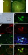
In vivo migration and differentiation of WA09 hESC-derived NCSCs. (A) DiO-labeled cells at time of injection and (B) 48 h later showing cell migration. (C–F) Immunocytochemistry and bright-field images of the same microscopic field 72 h after injection showing cells that had incorporated into a cranial ganglion area and differentiated to peripheral neurons. Cells were probed with antibodies for peripherin and hNA and counterstained with DAPI (DNA). (Scale bar, 50 μm.) (GJ) Images of the same microscopic field showing a cluster of human NCSCs (hNA-positive) in the head mesenchyme adjacent to the neural tube. Many of the cells are also Tuj1-positive. (Scale bar, 20 μm.)

References

    1. Selleck MA, Bronner-Fraser M. The genesis of avian neural crest cells: A classic embryonic induction. Proc Natl Acad Sci USA. 1996;93:9352–9357. - PMC - PubMed
    1. Meulemans D, Bronner-Fraser M. Gene-regulatory interactions in neural crest evolution and development. Dev Cell. 2004;7(3):291–299. - PubMed
    1. García-Castro MI, Marcelle C, Bronner-Fraser M. Ectodermal Wnt function as a neural crest inducer. Science. 2002;297:848–851. - PubMed
    1. Patthey C, Edlund T, Gunhaga L. Wnt-regulated temporal control of BMP exposure directs the choice between neural plate border and epidermal fate. Development. 2009;136(1):73–83. - PubMed
    1. Wilson SI, et al. The status of Wnt signalling regulates neural and epidermal fates in the chick embryo. Nature. 2001;411:325–330. - PubMed

Publication types

MeSH terms