

Analysis of pars plana vitrectomy for optic pit-related maculopathy with intraoperative optical coherence tomography: a possible connection with the vitreous cavity
- PMID: 22084218
- PMCID: PMC3509483
- DOI: 10.1001/archophthalmol.2011.316
Analysis of pars plana vitrectomy for optic pit-related maculopathy with intraoperative optical coherence tomography: a possible connection with the vitreous cavity
Abstract
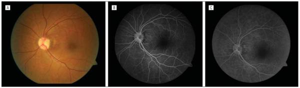
Optimal management of optic pit-related maculopathy remains to be determined. The fluid source for the maculopathy also remains controversial. In this article, we present a unique surgical technique for internal drainage of the intraretinal fluid and describe the intraoperative use of spectral-domain optical coherence tomography to assist in the surgical management of this condition. Pars plana vitrectomy was performed with elevation of the posterior hyaloid. Following an air-fluid exchange, aspiration over the optic nerve pit was performed. Following aspiration, intraoperative spectral-domain optical coherence tomography demonstrated collapse of the retinoschisis, strongly suggesting a connection between the vitreous cavity and the intraretinal fluid.
Figures

Comment in
-
Endodrainage of macular schisis through optic disc pit.Arch Ophthalmol. 2012 Jun;130(6):808-9. doi: 10.1001/archophthalmol.2012.10. Arch Ophthalmol. 2012. PMID: 22801858 No abstract available.
References
-
- Brown GC, Shields JA, Patty BE, Goldberg RE. Congenital pits of the optic nerve head, I: experimental studies in collie dogs. Arch Ophthalmol. 1979;97(7):1341–1344. - PubMed
-
- Gass JD. Serous detachment of the macula: secondary to congenital pit of the optic nervehead. Am J Ophthalmol. 1969;67(6):821–841. - PubMed
-
- Konno S, Akiba J, Sato E, Kuriyama S, Yoshida A. OCT in successful surgery of retinal detachment associated with optic nerve head pit. Ophthalmic Surg Lasers. 2000;31(3):236–239. - PubMed
-
- Rutledge BK, Puliafito CA, Duker JS, Hee MR, Cox MS. Optical coherence tomography of macular lesions associated with optic nerve head pits. Ophthalmology. 1996;103(7):1047–1053. - PubMed
Publication types
MeSH terms
Grants and funding
LinkOut - more resources
Full Text Sources
Other Literature Sources
Medical
Miscellaneous
